 Movie
Movie Controller

[English] 日本語
 Yorodumi
- PDB-3p8m: Human dynein light chain (DYNLL2) in complex with an in vitro evo... -
+ Open data

- Basic information
| Entry | Database: PDB / ID: 3p8m | ||||||
|---|---|---|---|---|---|---|---|
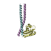
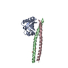
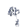
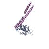
| Title | Human dynein light chain (DYNLL2) in complex with an in vitro evolved peptide dimerized by leucine zipper | ||||||
 Components Components |
| ||||||
 Keywords Keywords | PROTEIN BINDING / Phage display / Leucine zipper / Hub protein | ||||||
| Function / homology |  Function and homology information Function and homology informationmyosin V complex / Activation of BMF and translocation to mitochondria / 9+0 non-motile cilium / FCERI mediated MAPK activation / protein localization to nuclear periphery / Activation of the AP-1 family of transcription factors / negative regulation of ribosomal protein gene transcription by RNA polymerase II / positive regulation of cellular response to amino acid starvation / Intraflagellar transport / response to amino acid starvation ...myosin V complex / Activation of BMF and translocation to mitochondria / 9+0 non-motile cilium / FCERI mediated MAPK activation / protein localization to nuclear periphery / Activation of the AP-1 family of transcription factors / negative regulation of ribosomal protein gene transcription by RNA polymerase II / positive regulation of cellular response to amino acid starvation / Intraflagellar transport / response to amino acid starvation / mediator complex binding / Oxidative Stress Induced Senescence / COPI-independent Golgi-to-ER retrograde traffic / cytoplasmic dynein complex / Macroautophagy / dynein intermediate chain binding / amino acid biosynthetic process / TFIID-class transcription factor complex binding / positive regulation of RNA polymerase II transcription preinitiation complex assembly / positive regulation of transcription initiation by RNA polymerase II / microtubule-based process / COPI-mediated anterograde transport / ciliary tip / Amplification of signal from unattached kinetochores via a MAD2 inhibitory signal / MHC class II antigen presentation / HSP90 chaperone cycle for steroid hormone receptors (SHR) in the presence of ligand / cellular response to nutrient levels / Mitotic Prometaphase / EML4 and NUDC in mitotic spindle formation / cellular response to amino acid starvation / Resolution of Sister Chromatid Cohesion / RHO GTPases Activate Formins / RNA polymerase II transcription regulator complex / HCMV Early Events / Aggrephagy / Separation of Sister Chromatids / scaffold protein binding / DNA-binding transcription activator activity, RNA polymerase II-specific / transcription regulator complex / sequence-specific DNA binding / microtubule / RNA polymerase II-specific DNA-binding transcription factor binding / cytoskeleton / DNA-binding transcription factor activity, RNA polymerase II-specific / cilium / postsynapse / postsynaptic density / intracellular signal transduction / RNA polymerase II cis-regulatory region sequence-specific DNA binding / DNA-binding transcription factor activity / chromatin binding / centrosome / protein-containing complex binding / glutamatergic synapse / negative regulation of transcription by RNA polymerase II / positive regulation of transcription by RNA polymerase II / membrane / identical protein binding / nucleus / plasma membrane / cytosol Similarity search - Function | ||||||
| Biological species |  Homo sapiens (human) Homo sapiens (human) | ||||||
| Method |  X-RAY DIFFRACTION / MOLECULAR REPLACEMENT / Resolution: 2.9 Å X-RAY DIFFRACTION / MOLECULAR REPLACEMENT / Resolution: 2.9 Å | ||||||
 Authors Authors | Rapali, P. / Radnai, L. / Suveges, D. / Hetenyi, C. / Harmat, V. / Tolgyesi, F. / Wahlgren, W.Y. / Katona, G. / Nyitray, L. / Pal, G. | ||||||
 Citation Citation | Journal: Plos One / Year: 2011 Title: Directed evolution reveals the binding motif preference of the LC8/DYNLL hub protein and predicts large numbers of novel binders in the human proteome. Authors: Rapali, P. / Radnai, L. / Suveges, D. / Harmat, V. / Tolgyesi, F. / Wahlgren, W.Y. / Katona, G. / Nyitray, L. / Pal, G. | ||||||
| History |
|
- Structure visualization
| Structure viewer | Molecule: MolmilJmol/JSmol |
|---|
- Downloads & links
-Download
| PDBx/mmCIF format | 3p8m.cif.gz | 116.9 KB | Display | PDBx/mmCIF format |
|---|---|---|---|---|
| PDB format | pdb3p8m.ent.gz | 91.6 KB | Display | PDB format |
| PDBx/mmJSON format | 3p8m.json.gz | Tree view | PDBx/mmJSON format | |
| Others |  Other downloads Other downloads |
-Validation report
| Arichive directory | https://data.pdbj.org/pub/pdb/validation_reports/p8/3p8mftp://data.pdbj.org/pub/pdb/validation_reports/p8/3p8m | HTTPS FTP |
|---|
-Related structure data
| Related structure data |  2xqqSC S: Starting model for refinement C: citing same article ( |
|---|---|
| Similar structure data |


-Links
 PDBj
PDBj


- Assembly
| Deposited unit | 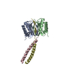
| |||||||||||||||||||||||||||||||||||||||||||||||||||||||||||||||||||||||||||||||||||||||||||||||
|---|---|---|---|---|---|---|---|---|---|---|---|---|---|---|---|---|---|---|---|---|---|---|---|---|---|---|---|---|---|---|---|---|---|---|---|---|---|---|---|---|---|---|---|---|---|---|---|---|---|---|---|---|---|---|---|---|---|---|---|---|---|---|---|---|---|---|---|---|---|---|---|---|---|---|---|---|---|---|---|---|---|---|---|---|---|---|---|---|---|---|---|---|---|---|---|---|
| 1 |
| |||||||||||||||||||||||||||||||||||||||||||||||||||||||||||||||||||||||||||||||||||||||||||||||
| Unit cell |
| |||||||||||||||||||||||||||||||||||||||||||||||||||||||||||||||||||||||||||||||||||||||||||||||
| Noncrystallographic symmetry (NCS) | NCS domain:
NCS domain segments: Component-ID: 1 / Refine code: 3
NCS ensembles :
|
-Components
| #1: Protein | Mass: 10647.124 Da / Num. of mol.: 2 Source method: isolated from a genetically manipulated source Source: (gene. exp.) Homo sapiens (human) / Gene: DYNLL2, DLC2 / Plasmid: pET-15b / Production host:  #2: Protein/peptide | Mass: 5110.736 Da / Num. of mol.: 2 Source method: isolated from a genetically manipulated source Source: (gene. exp.) Gene: GCN4, AAS3, ARG9, YEL009C / Plasmid: modified pET / Production host: #3: Water | ChemComp-HOH / |  Mass: 18.015 Da / Num. of mol.: 41 / Source method: isolated from a natural source / Formula: H2O Mass: 18.015 Da / Num. of mol.: 41 / Source method: isolated from a natural source / Formula: H2OSequence details | THE IN VITRO EVOLVED BINDING SEQUENCE. | |
|---|
-Experimental details
-Experiment
| Experiment | Method: X-RAY DIFFRACTION / Number of used crystals: 1 |
|---|
- Sample preparation
| Crystal | Density Matthews: 2.97 Å3/Da / Density % sol: 58.6 % |
|---|---|
| Crystal grow | Temperature: 293 K / pH: 7 Details: 20% PEG8000, 0.2 M MgCl2, 0.1 M TRIS, pH 7.0, VAPOR DIFFUSION, HANGING DROP, temperature 293K |
-Data collection
| Diffraction | Mean temperature: 100 K |
|---|---|
| Diffraction source | Source: ROTATING ANODE / Type: RIGAKU RUH2R / Wavelength: 1.5418 |
| Detector | Type: RIGAKU RAXIS IV++ / Detector: IMAGE PLATE / Date: Mar 1, 2010 / Details: OSMIC BLUE |
| Radiation | Monochromator: CONFOCAL MIRRORS / Protocol: SINGLE WAVELENGTH / Monochromatic (M) / Laue (L): M / Scattering type: x-ray |
| Radiation wavelength | Wavelength: 1.5418 Å / Relative weight: 1 |
| Reflection | Resolution: 2.9→56.76 Å / Num. obs: 8273 / % possible obs: 94.4 % / Observed criterion σ(I): -3 / Redundancy: 3.23 % / Rmerge(I) obs: 0.131 / Net I/σ(I): 6.4 |
| Reflection shell | Resolution: 2.9→3 Å / Redundancy: 3.12 % / Rmerge(I) obs: 0.576 / Mean I/σ(I) obs: 1.5 / % possible all: 96.6 |
- Processing
| Software |
| |||||||||||||||||||||||||||||||||||||||||||||||||||||||||||||||||||||||||||||||||||||||||||||||||||||||||||||||||||||||||||||||||||||||||||||||||||||||||||||||||||||||||||||||
|---|---|---|---|---|---|---|---|---|---|---|---|---|---|---|---|---|---|---|---|---|---|---|---|---|---|---|---|---|---|---|---|---|---|---|---|---|---|---|---|---|---|---|---|---|---|---|---|---|---|---|---|---|---|---|---|---|---|---|---|---|---|---|---|---|---|---|---|---|---|---|---|---|---|---|---|---|---|---|---|---|---|---|---|---|---|---|---|---|---|---|---|---|---|---|---|---|---|---|---|---|---|---|---|---|---|---|---|---|---|---|---|---|---|---|---|---|---|---|---|---|---|---|---|---|---|---|---|---|---|---|---|---|---|---|---|---|---|---|---|---|---|---|---|---|---|---|---|---|---|---|---|---|---|---|---|---|---|---|---|---|---|---|---|---|---|---|---|---|---|---|---|---|---|---|---|---|
| Refinement | Method to determine structure: MOLECULAR REPLACEMENT Starting model: PDB ENTRY 2XQQ Resolution: 2.9→42.31 Å / Cor.coef. Fo:Fc: 0.915 / Cor.coef. Fo:Fc free: 0.894 / SU B: 46.408 / SU ML: 0.391 / Cross valid method: THROUGHOUT / σ(F): 0 / ESU R Free: 0.451 / Stereochemistry target values: MAXIMUM LIKELIHOOD / Details: HYDROGENS HAVE BEEN ADDED IN THE RIDING POSITIONS
| |||||||||||||||||||||||||||||||||||||||||||||||||||||||||||||||||||||||||||||||||||||||||||||||||||||||||||||||||||||||||||||||||||||||||||||||||||||||||||||||||||||||||||||||
| Solvent computation | Ion probe radii: 0.8 Å / Shrinkage radii: 0.8 Å / VDW probe radii: 1.4 Å / Solvent model: BABINET MODEL WITH MASK | |||||||||||||||||||||||||||||||||||||||||||||||||||||||||||||||||||||||||||||||||||||||||||||||||||||||||||||||||||||||||||||||||||||||||||||||||||||||||||||||||||||||||||||||
| Displacement parameters | Biso mean: 52.8 Å2
| |||||||||||||||||||||||||||||||||||||||||||||||||||||||||||||||||||||||||||||||||||||||||||||||||||||||||||||||||||||||||||||||||||||||||||||||||||||||||||||||||||||||||||||||
| Refinement step | Cycle: LAST / Resolution: 2.9→42.31 Å
| |||||||||||||||||||||||||||||||||||||||||||||||||||||||||||||||||||||||||||||||||||||||||||||||||||||||||||||||||||||||||||||||||||||||||||||||||||||||||||||||||||||||||||||||
| Refine LS restraints |
| |||||||||||||||||||||||||||||||||||||||||||||||||||||||||||||||||||||||||||||||||||||||||||||||||||||||||||||||||||||||||||||||||||||||||||||||||||||||||||||||||||||||||||||||
| Refine LS restraints NCS | Refine-ID: X-RAY DIFFRACTION
| |||||||||||||||||||||||||||||||||||||||||||||||||||||||||||||||||||||||||||||||||||||||||||||||||||||||||||||||||||||||||||||||||||||||||||||||||||||||||||||||||||||||||||||||
| LS refinement shell | Resolution: 2.9→2.98 Å / Total num. of bins used: 20
| |||||||||||||||||||||||||||||||||||||||||||||||||||||||||||||||||||||||||||||||||||||||||||||||||||||||||||||||||||||||||||||||||||||||||||||||||||||||||||||||||||||||||||||||
| Refinement TLS params. | Method: refined / Refine-ID: X-RAY DIFFRACTION
| |||||||||||||||||||||||||||||||||||||||||||||||||||||||||||||||||||||||||||||||||||||||||||||||||||||||||||||||||||||||||||||||||||||||||||||||||||||||||||||||||||||||||||||||
| Refinement TLS group |
|
