[English] 日本語
Yorodumi
- PDB-3p0i: Leishmania major Tyrosyl-tRNA synthetase in complex with tyrosino... -

+
Open data


ID or keywords:

Loading...

-
Basic information

Entry
Database: PDB / ID: 3p0i
TitleLeishmania major Tyrosyl-tRNA synthetase in complex with tyrosinol, cubic crystal form
ComponentsTyrosyl-tRNA synthetase
KeywordsLIGASE / aminoacyl-tRNA synthetase / tRNA ligase / aaRS / TyrRS / pseudodimer / translation / ATP-binding / nucleotide-binding / Structural Genomics / Medical Structural Genomics of Pathogenic Protozoa / MSGPP
Function / homology
Function and homology information


tyrosyl-tRNA aminoacylation / tyrosine-tRNA ligase / tyrosine-tRNA ligase activity / ciliary plasm / ATP binding / cytoplasm
Similarity search - Function
Tyrosyl-Transfer RNA Synthetase / Tyrosyl-Transfer RNA Synthetase / Aminoacyl-tRNA synthetase, class Ic / tRNA synthetases class I (W and Y) / HUPs / Rossmann-like alpha/beta/alpha sandwich fold / Rossmann fold / Orthogonal Bundle / 3-Layer(aba) Sandwich / Mainly Alpha / Alpha Beta
Similarity search - Domain/homology
4-[(2S)-2-amino-3-hydroxypropyl]phenol / tyrosine--tRNA ligase
Similarity search - Component
Biological speciesLeishmania major (eukaryote)
MethodX-RAY DIFFRACTION / SYNCHROTRON / FOURIER SYNTHESIS / molecular replacement / Resolution: 3.13 Å
AuthorsMerritt, E.A. / Larson, E.T. / Medical Structural Genomics of Pathogenic Protozoa (MSGPP)
CitationJournal: J.Mol.Biol. / Year: 2011
Title: The Double-Length Tyrosyl-tRNA Synthetase from the Eukaryote Leishmania major Forms an Intrinsically Asymmetric Pseudo-Dimer.
Authors: Larson, E.T. / Kim, J.E. / Castaneda, L.J. / Napuli, A.J. / Zhang, Z. / Fan, E. / Zucker, F.H. / Verlinde, C.L. / Buckner, F.S. / Van Voorhis, W.C. / Hol, W.G. / Merritt, E.A.
History
DepositionSep 28, 2010Deposition site: RCSB / Processing site: RCSB
Revision 1.0Mar 23, 2011Provider: repository / Type: Initial release
Revision 1.1Jul 13, 2011Group: Version format compliance
Revision 1.2Sep 6, 2023Group: Data collection / Database references ...Data collection / Database references / Derived calculations / Refinement description / Structure summary
Category: chem_comp / chem_comp_atom ...chem_comp / chem_comp_atom / chem_comp_bond / database_2 / pdbx_initial_refinement_model / struct_conn / struct_ref_seq_dif / struct_site
Item: _chem_comp.mon_nstd_flag / _chem_comp.pdbx_synonyms ..._chem_comp.mon_nstd_flag / _chem_comp.pdbx_synonyms / _chem_comp.type / _database_2.pdbx_DOI / _database_2.pdbx_database_accession / _struct_conn.pdbx_leaving_atom_flag / _struct_ref_seq_dif.details / _struct_site.pdbx_auth_asym_id / _struct_site.pdbx_auth_comp_id / _struct_site.pdbx_auth_seq_id
Revision 1.3Dec 6, 2023Group: Data collection / Category: chem_comp_atom / chem_comp_bond / Item: _chem_comp_atom.atom_id / _chem_comp_bond.atom_id_2

-
Structure visualization

Structure viewerMolecule:
MolmilJmol/JSmol

Downloads & links

-
Assembly

Deposited unit
A: Tyrosyl-tRNA synthetase
B: Tyrosyl-tRNA synthetase
hetero molecules


Theoretical massNumber of molelcules
Total (without water)154,4494
Polymers154,1142
Non-polymers3342
Water0
1
A: Tyrosyl-tRNA synthetase
hetero molecules


Theoretical massNumber of molelcules
Total (without water)77,2242
Polymers77,0571
Non-polymers1671
Water0
TypeNameSymmetry operationNumber
identity operation1_555x,y,z1
MethodPISA
2
B: Tyrosyl-tRNA synthetase
hetero molecules


Theoretical massNumber of molelcules
Total (without water)77,2242
Polymers77,0571
Non-polymers1671
Water0
TypeNameSymmetry operationNumber
identity operation1_555x,y,z1
MethodPISA
Unit cell
Length a, b, c (Å)241.400, 241.400, 241.400
Angle α, β, γ (deg.)90.00, 90.00, 90.00
Int Tables number197
Space group name H-MI23
Noncrystallographic symmetry (NCS)NCS domain:
IDEns-IDDetails
11A
21B
12A
22B

NCS domain segments:
Dom-IDComponent-IDEns-IDRefine codeAuth asym-IDAuth seq-ID
1112A0 - 363
2112B0 - 363
1122A365 - 682
2122B365 - 682

NCS ensembles :
ID
1
2

-
Components

#1: Protein Tyrosyl-tRNA synthetase


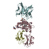
Mass: 77057.195 Da / Num. of mol.: 2
Source method: isolated from a genetically manipulated source
Source: (gene. exp.) Leishmania major (eukaryote) / Gene: LmjF14.1370 / Plasmid: BG1861 / Production host: Escherichia coli (E. coli) / Strain (production host): BL21(DE3) / References: UniProt: Q4QFJ7, tyrosine-tRNA ligase
#2: Chemical ChemComp-TYE / 4-[(2S)-2-amino-3-hydroxypropyl]phenol / tyrosinol / bound form of TYROSINAL / Tyrosinol


Type: L-peptide linking / Mass: 167.205 Da / Num. of mol.: 2 / Source method: obtained synthetically / Formula: C9H13NO2

-
Experimental details

-
Experiment

ExperimentMethod: X-RAY DIFFRACTION / Number of used crystals: 1

-
Sample preparation

CrystalDensity Matthews: 3.8 Å3/Da / Density % sol: 67.66 %
Crystal growTemperature: 277 K / Method: vapor diffusion, sitting drop
Details: equal volumes SeMet protein solution (24 mg/ml LmTyrRS in MSGPP buffer containing 5 mM DTT and 10 mM tyrosinol) and well solution (25% PEG 3350, 0.1 M tri-sodium citrate pH 5.5, 10 mM Ferric ...Details: equal volumes SeMet protein solution (24 mg/ml LmTyrRS in MSGPP buffer containing 5 mM DTT and 10 mM tyrosinol) and well solution (25% PEG 3350, 0.1 M tri-sodium citrate pH 5.5, 10 mM Ferric III Chloride); cryoprotected by addition of 26% PEG 3350, 8% MSGPP buffer, 8% ethylene glycol, 20% glycerol and 0.09 M tri-sodium citrate pH 5.5 directly to drop prior to mounting and freezing crystals in liquid nitrogen, VAPOR DIFFUSION, SITTING DROP, temperature 277K

-
Data collection

DiffractionMean temperature: 100 K
Diffraction sourceSource: SYNCHROTRON / Site: SSRL / Beamline: BL9-2 / Wavelength: 0.97908 Å
DetectorType: MARMOSAIC 325 mm CCD / Detector: CCD / Date: Mar 19, 2010
RadiationMonochromator: crystal / Protocol: SINGLE WAVELENGTH / Monochromatic (M) / Laue (L): M / Scattering type: x-ray
Radiation wavelengthWavelength: 0.97908 Å / Relative weight: 1
ReflectionResolution: 3.13→85.35 Å / Num. obs: 41253 / % possible obs: 99.93 % / Redundancy: 7.98 % / Biso Wilson estimate: 83 Å2 / Rmerge(I) obs: 0.182 / Net I/σ(I): 7.3331
Reflection shell
Resolution (Å)Redundancy (%)Rmerge(I) obsDiffraction-ID% possible all
3.13-3.36.960.014199.82
3.3-3.57.340.014199.92
3.5-3.747.590.014199.91
3.74-4.047.860.014199.95
4.04-4.428.360.0141100
4.42-4.958.750.0141100
4.95-5.718.790.0141100
5.71-78.760.0141100
7-9.898.670.0141100
9.89-85.358.260.014199.52

-
Phasing

PhasingMethod: molecular replacement

-
Processing

Software
NameVersionClassificationNB
MOSFLMdata reduction
SCALACCP4_3.3.16data scaling
REFMACrefmac_5.5.0110refinement
PDB_EXTRACT3.1data extraction
Blu-Icedata collection
REFMACphasing
RefinementMethod to determine structure: FOURIER SYNTHESIS
Starting model: PDB entry 3P0H
Resolution: 3.13→85.35 Å / Cor.coef. Fo:Fc: 0.911 / Cor.coef. Fo:Fc free: 0.898 / Occupancy max: 1 / Occupancy min: 0.5 / SU B: 48.246 / SU ML: 0.369 / Isotropic thermal model: overall / Cross valid method: THROUGHOUT / σ(F): 0 / ESU R Free: 0.416 / Stereochemistry target values: MAXIMUM LIKELIHOOD
Details: HYDROGENS HAVE BEEN ADDED IN THE RIDING POSITIONS. U VALUES RESIDUAL ONLY.
RfactorNum. reflection% reflectionSelection details
Rfree0.2679 2062 5 %RANDOM
Rwork0.2395 ---
obs0.2409 41212 99.81 %-
Solvent computationIon probe radii: 0.8 Å / Shrinkage radii: 0.8 Å / VDW probe radii: 1.4 Å / Solvent model: MASK
Displacement parametersBiso mean: 122.862 Å2
Refinement stepCycle: LAST / Resolution: 3.13→85.35 Å
ProteinNucleic acidLigandSolventTotal
Num. atoms9278 0 24 0 9302
Refine LS restraints
Refine-IDTypeDev idealDev ideal targetNumber
X-RAY DIFFRACTIONr_bond_refined_d0.010.0229476
X-RAY DIFFRACTIONr_bond_other_d0.0020.026315
X-RAY DIFFRACTIONr_angle_refined_deg1.1621.96412845
X-RAY DIFFRACTIONr_angle_other_deg0.925315432
X-RAY DIFFRACTIONr_dihedral_angle_1_deg5.94551207
X-RAY DIFFRACTIONr_dihedral_angle_2_deg36.92124.458406
X-RAY DIFFRACTIONr_dihedral_angle_3_deg18.697151637
X-RAY DIFFRACTIONr_dihedral_angle_4_deg19.7181558
X-RAY DIFFRACTIONr_chiral_restr0.0650.21479
X-RAY DIFFRACTIONr_gen_planes_refined0.0050.0210563
X-RAY DIFFRACTIONr_gen_planes_other0.0010.021819
X-RAY DIFFRACTIONr_nbd_refined
X-RAY DIFFRACTIONr_nbd_other
X-RAY DIFFRACTIONr_nbtor_refined
X-RAY DIFFRACTIONr_nbtor_other
X-RAY DIFFRACTIONr_xyhbond_nbd_refined
X-RAY DIFFRACTIONr_xyhbond_nbd_other
X-RAY DIFFRACTIONr_metal_ion_refined
X-RAY DIFFRACTIONr_metal_ion_other
X-RAY DIFFRACTIONr_symmetry_vdw_refined
X-RAY DIFFRACTIONr_symmetry_vdw_other
X-RAY DIFFRACTIONr_symmetry_hbond_refined
X-RAY DIFFRACTIONr_symmetry_hbond_other
X-RAY DIFFRACTIONr_symmetry_metal_ion_refined
X-RAY DIFFRACTIONr_symmetry_metal_ion_other
X-RAY DIFFRACTIONr_mcbond_it
X-RAY DIFFRACTIONr_mcbond_other
X-RAY DIFFRACTIONr_mcangle_it
X-RAY DIFFRACTIONr_scbond_it
X-RAY DIFFRACTIONr_scangle_it
X-RAY DIFFRACTIONr_rigid_bond_restr
X-RAY DIFFRACTIONr_sphericity_free
X-RAY DIFFRACTIONr_sphericity_bonded
Refine LS restraints NCS

Dom-ID: 1 / Auth asym-ID: A / Refine-ID: X-RAY DIFFRACTION

Ens-IDNumberTypeRms dev position (Å)Weight position
12105TIGHT POSITIONAL0.020.05
12581MEDIUM POSITIONAL0.030.5
21056TIGHT POSITIONAL0.020.05
21148MEDIUM POSITIONAL0.020.5
LS refinement shellResolution: 3.13→3.21 Å / Total num. of bins used: 20
RfactorNum. reflection% reflection
Rfree0.362 165 -
Rwork0.35 2833 -
all-2998 -
obs--99.54 %
Refinement TLS params.

Method: refined / Refine-ID: X-RAY DIFFRACTION

IDL112)L122)L132)L222)L232)L332)S11 (Å °)S12 (Å °)S13 (Å °)S21 (Å °)S22 (Å °)S23 (Å °)S31 (Å °)S32 (Å °)S33 (Å °)T112)T122)T132)T222)T232)T332)Origin x (Å)Origin y (Å)Origin z (Å)
16.38832.20071.80772.3293-0.2331.31810.0694-0.4957-0.48640.4547-0.1708-0.19570.0503-0.04940.10150.31930.13650.16060.25530.16590.531922.41267.37764.14
28.44222.8698-2.54242.9094-0.54552.66280.18040.28170.03910.0944-0.10360.63080.3889-0.3141-0.07680.4477-0.05320.12950.29390.07150.8158-24.44646.38169.548
32.8995-1.4522-0.82687.03251.62051.6380.00460.54590.2811-0.4573-0.0070.0338-0.349-0.19080.00240.19960.13650.05630.35360.11480.50823.72892.32841.417
416.2024-3.7914-11.34823.82363.508411.04220.59513.5367-0.4229-0.578-0.90890.4050.2049-2.41850.31380.46360.15160.01831.4473-0.18510.642719.9869.50210.545
Refinement TLS group
IDRefine-IDRefine TLS-IDAuth asym-IDAuth seq-ID
1X-RAY DIFFRACTION1A-1 - 364
2X-RAY DIFFRACTION1A701
3X-RAY DIFFRACTION2A365 - 680
4X-RAY DIFFRACTION3B-1 - 355
5X-RAY DIFFRACTION3B701
6X-RAY DIFFRACTION4B366 - 544

+
About Yorodumi

-
News

-
Feb 9, 2022. New format data for meta-information of EMDB entries

New format data for meta-information of EMDB entries

  • Version 3 of the EMDB header file is now the official format.
  • The previous official version 1.9 will be removed from the archive.

Related info.:EMDB header

External links:wwPDB to switch to version 3 of the EMDB data model

-
Aug 12, 2020. Covid-19 info

Covid-19 info

URL: https://pdbj.org/emnavi/covid19.php

New page: Covid-19 featured information page in EM Navigator.

Related info.:Covid-19 info / Mar 5, 2020. Novel coronavirus structure data

+
Mar 5, 2020. Novel coronavirus structure data

Novel coronavirus structure data

Related info.:Yorodumi Speices / Aug 12, 2020. Covid-19 info

External links:COVID-19 featured content - PDBj / Molecule of the Month (242):Coronavirus Proteases

+
Jan 31, 2019. EMDB accession codes are about to change! (news from PDBe EMDB page)

EMDB accession codes are about to change! (news from PDBe EMDB page)

  • The allocation of 4 digits for EMDB accession codes will soon come to an end. Whilst these codes will remain in use, new EMDB accession codes will include an additional digit and will expand incrementally as the available range of codes is exhausted. The current 4-digit format prefixed with “EMD-” (i.e. EMD-XXXX) will advance to a 5-digit format (i.e. EMD-XXXXX), and so on. It is currently estimated that the 4-digit codes will be depleted around Spring 2019, at which point the 5-digit format will come into force.
  • The EM Navigator/Yorodumi systems omit the EMD- prefix.

Related info.:Q: What is EMD? / ID/Accession-code notation in Yorodumi/EM Navigator

External links:EMDB Accession Codes are Changing Soon! / Contact to PDBj

+
Jul 12, 2017. Major update of PDB

Major update of PDB

  • wwPDB released updated PDB data conforming to the new PDBx/mmCIF dictionary.
  • This is a major update changing the version number from 4 to 5, and with Remediation, in which all the entries are updated.
  • In this update, many items about electron microscopy experimental information are reorganized (e.g. em_software).
  • Now, EM Navigator and Yorodumi are based on the updated data.

External links:wwPDB Remediation / Enriched Model Files Conforming to OneDep Data Standards Now Available in the PDB FTP Archive

-
Yorodumi

Thousand views of thousand structures

  • Yorodumi is a browser for structure data from EMDB, PDB, SASBDB, etc.
  • This page is also the successor to EM Navigator detail page, and also detail information page/front-end page for Omokage search.
  • The word "yorodu" (or yorozu) is an old Japanese word meaning "ten thousand". "mi" (miru) is to see.

Related info.:EMDB / PDB / SASBDB / Comparison of 3 databanks / Yorodumi Search / Aug 31, 2016. New EM Navigator & Yorodumi / Yorodumi Papers / Jmol/JSmol / Function and homology information / Changes in new EM Navigator and Yorodumi

Read more