 Movie
Movie Controller


+ Open data

- Basic information
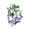
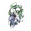
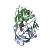
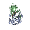
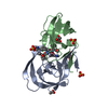
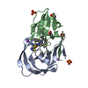
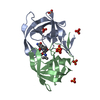
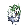
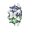
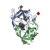
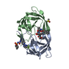
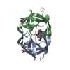
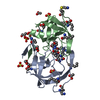
| Entry | Database: PDB / ID: 3oxc | |||||||||
|---|---|---|---|---|---|---|---|---|---|---|
| Title | Wild Type HIV-1 Protease with Antiviral Drug Saquinavir | |||||||||
 Components Components | Protease | |||||||||
 Keywords Keywords | HYDROLASE/HYDROLASE INHIBITOR / enzyme inhibition / aspartic protease / HIV/AIDS / Saquinavir / HYDROLASE-HYDROLASE INHIBITOR complex | |||||||||
| Function / homology |  Function and homology information Function and homology informationhost multivesicular body / aspartic-type endopeptidase activity / virion membrane / proteolysis / identical protein binding Similarity search - Function | |||||||||
| Biological species |   Human immunodeficiency virus 1 Human immunodeficiency virus 1 | |||||||||
| Method |  X-RAY DIFFRACTION / SYNCHROTRON / MOLECULAR REPLACEMENT / Resolution: 1.16 Å X-RAY DIFFRACTION / SYNCHROTRON / MOLECULAR REPLACEMENT / Resolution: 1.16 Å | |||||||||
 Authors Authors | Kovalevsky, A.Y. / Wang, Y.-F. / Tie, Y. / Weber, I.T. | |||||||||
 Citation Citation | Journal: Proteins / Year: 2007 Title: Atomic resolution crystal structures of HIV-1 protease and mutants V82A and I84V with saquinavir Authors: Kovalevsky, A.Y. / Wang, Y.-F. / Tie, Y. / Weber, I.T. #1: Journal: To be PublishedAuthors: Tie, Y. / Kovalevsky, A.Y. / Boross, P. / Wang, Y.-F. / Ghosh, A.K. / Tozser, T. / Harrison, R.W. / Weber, I.T. | |||||||||
| History |
|
- Structure visualization
| Structure viewer | Molecule: MolmilJmol/JSmol |
|---|
- Downloads & links
-Download
| PDBx/mmCIF format | 3oxc.cif.gz | 108.8 KB | Display | PDBx/mmCIF format |
|---|---|---|---|---|
| PDB format | pdb3oxc.ent.gz | 82.8 KB | Display | PDB format |
| PDBx/mmJSON format | 3oxc.json.gz | Tree view | PDBx/mmJSON format | |
| Others |  Other downloads Other downloads |
-Validation report
| Arichive directory | https://data.pdbj.org/pub/pdb/validation_reports/ox/3oxcftp://data.pdbj.org/pub/pdb/validation_reports/ox/3oxc | HTTPS FTP |
|---|
-Related structure data
| Related structure data | 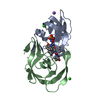 2nmySC  2nmzC 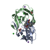 2nnkC 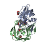 2nnpC S: Starting model for refinement C: citing same article ( |
|---|---|
| Similar structure data |


-Links
 PDBj
PDBj
- Assembly
| Deposited unit | 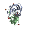
| ||||||||
|---|---|---|---|---|---|---|---|---|---|
| 1 |
| ||||||||
| Unit cell |
|
-Components
| #1: Protein | Mass: 10740.677 Da / Num. of mol.: 2 / Fragment: residues 500-598 / Mutation: Q7K, L33I, L63I, C67A, C95A Source method: isolated from a genetically manipulated source Source: (gene. exp.) Human immunodeficiency virus 1 / Gene: pol / Plasmid: pET11a / Production host:  #2: Chemical | ChemComp-ROC / ( |   Type: peptide-like, Peptide-like / Class: Inhibitor / Mass: 670.841 Da / Num. of mol.: 1 / Source method: obtained synthetically / Formula: C38H50N6O5 / References: Saquinavir / Comment: medication, antiretroviral*YM Type: peptide-like, Peptide-like / Class: Inhibitor / Mass: 670.841 Da / Num. of mol.: 1 / Source method: obtained synthetically / Formula: C38H50N6O5 / References: Saquinavir / Comment: medication, antiretroviral*YM#3: Chemical |   Mass: 46.025 Da / Num. of mol.: 2 / Source method: obtained synthetically / Formula: CH2O2 Mass: 46.025 Da / Num. of mol.: 2 / Source method: obtained synthetically / Formula: CH2O2#4: Chemical |   Mass: 96.063 Da / Num. of mol.: 2 / Source method: obtained synthetically / Formula: SO4 Mass: 96.063 Da / Num. of mol.: 2 / Source method: obtained synthetically / Formula: SO4#5: Water | ChemComp-HOH / |  Mass: 18.015 Da / Num. of mol.: 201 / Source method: isolated from a natural source / Formula: H2O Mass: 18.015 Da / Num. of mol.: 201 / Source method: isolated from a natural source / Formula: H2O |
|---|
-Experimental details
-Experiment
| Experiment | Method: X-RAY DIFFRACTION / Number of used crystals: 1 |
|---|
- Sample preparation
| Crystal | Density Matthews: 2.27 Å3/Da / Density % sol: 45.7 % |
|---|---|
| Crystal grow | Temperature: 298 K / Method: vapor diffusion, hanging drop / pH: 5.4 Details: AMMONIUM SULFATE 40%, CITRATE PHOSPHATE BUFFER PH 5.4, VAPOR DIFFUSION, HANGING DROP, temperature 298 K |
-Data collection
| Diffraction | Mean temperature: 90 K |
|---|---|
| Diffraction source | Source: SYNCHROTRON / Site: APS  / Beamline: 22-ID / Wavelength: 1 Å / Beamline: 22-ID / Wavelength: 1 Å |
| Detector | Type: MARMOSAIC 300 mm CCD / Detector: CCD / Date: Nov 11, 2004 |
| Radiation | Protocol: SINGLE WAVELENGTH / Monochromatic (M) / Laue (L): M / Scattering type: x-ray |
| Radiation wavelength | Wavelength: 1 Å / Relative weight: 1 |
| Reflection | Resolution: 1.16→50 Å / Num. all: 67949 / Num. obs: 67949 / % possible obs: 96.6 % / Redundancy: 5.6 % / Biso Wilson estimate: 9.946 Å2 / Rmerge(I) obs: 0.075 / Net I/σ(I): 14.6 |
| Reflection shell | Resolution: 1.16→1.2 Å / Redundancy: 3 % / Rmerge(I) obs: 0.348 / Mean I/σ(I) obs: 3 / % possible all: 72.6 |
- Processing
| Software |
| |||||||||||||||||||||||||||||||||
|---|---|---|---|---|---|---|---|---|---|---|---|---|---|---|---|---|---|---|---|---|---|---|---|---|---|---|---|---|---|---|---|---|---|---|
| Refinement | Method to determine structure: MOLECULAR REPLACEMENT Starting model: 2NMY Resolution: 1.16→10 Å / Num. parameters: 17373 / Num. restraintsaints: 22690 / Cross valid method: FREE R / σ(F): 0 / Stereochemistry target values: Engh & Huber / Details: ANISOTROPIC REFINEMENT REDUCED FREE R (NO CUTOFF)
| |||||||||||||||||||||||||||||||||
| Refine analyze | Num. disordered residues: 24 / Occupancy sum hydrogen: 1640.7 / Occupancy sum non hydrogen: 1762.9 | |||||||||||||||||||||||||||||||||
| Refinement step | Cycle: LAST / Resolution: 1.16→10 Å
| |||||||||||||||||||||||||||||||||
| Refine LS restraints |
|
