 Movie
Movie Controller


+ Open data

- Basic information
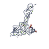
| Entry | Database: PDB / ID: 3ox0 | ||||||
|---|---|---|---|---|---|---|---|
| Title | Crystal structure of glycine riboswitch, unbound state | ||||||
 Components Components | (Domain II of glycine riboswitch) x 2 | ||||||
 Keywords Keywords | RNA / gene expression regulator / glycine riboswitch | ||||||
| Function / homology | : / RNA / RNA (> 10) Function and homology information Function and homology information | ||||||
| Method |  X-RAY DIFFRACTION / SYNCHROTRON / MOLECULAR REPLACEMENT / Resolution: 3.049 Å X-RAY DIFFRACTION / SYNCHROTRON / MOLECULAR REPLACEMENT / Resolution: 3.049 Å | ||||||
 Authors Authors | Huang, L. / Serganov, A. / Patel, D.J. | ||||||
 Citation Citation | Journal: Mol.Cell / Year: 2010 Title: Structural insights into ligand recognition by a sensing domain of the cooperative glycine riboswitch. Authors: Huang, L. / Serganov, A. / Patel, D.J. | ||||||
| History |
|
- Structure visualization
| Structure viewer | Molecule: MolmilJmol/JSmol |
|---|
- Downloads & links
-Download
| PDBx/mmCIF format | 3ox0.cif.gz | 205.4 KB | Display | PDBx/mmCIF format |
|---|---|---|---|---|
| PDB format | pdb3ox0.ent.gz | 164.4 KB | Display | PDB format |
| PDBx/mmJSON format | 3ox0.json.gz | Tree view | PDBx/mmJSON format | |
| Others |  Other downloads Other downloads |
-Validation report
| Arichive directory | https://data.pdbj.org/pub/pdb/validation_reports/ox/3ox0ftp://data.pdbj.org/pub/pdb/validation_reports/ox/3ox0 | HTTPS FTP |
|---|
-Related structure data
| Related structure data |  3owiSC  3owwC  3owzC 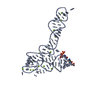 3oxbC  3oxdC 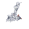 3oxeC  3oxjC  3oxmC S: Starting model for refinement C: citing same article ( |
|---|---|
| Similar structure data |


-Links
 PDBj
PDBj


- Assembly
| Deposited unit | 
| ||||||||
|---|---|---|---|---|---|---|---|---|---|
| 1 | 
| ||||||||
| 2 | 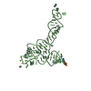
| ||||||||
| 3 |
| ||||||||
| Unit cell |
|
-Components
| #1: RNA chain | Mass: 28735.090 Da / Num. of mol.: 1 / Source method: obtained synthetically / Details: in vitro transcription / References: GenBank: CP001485.1 | ||||
|---|---|---|---|---|---|
| #2: RNA chain | Mass: 28655.109 Da / Num. of mol.: 1 / Source method: obtained synthetically / Details: in vitro transcription / References: GenBank: CP001485.1 | ||||
| #3: Chemical | ChemComp-MG /   Mass: 24.305 Da / Num. of mol.: 28 / Source method: obtained synthetically / Formula: Mg Mass: 24.305 Da / Num. of mol.: 28 / Source method: obtained synthetically / Formula: Mg#4: Water | ChemComp-HOH / |  Mass: 18.015 Da / Num. of mol.: 64 / Source method: isolated from a natural source / Formula: H2O Mass: 18.015 Da / Num. of mol.: 64 / Source method: isolated from a natural source / Formula: H2ONonpolymer details | THE ORIGINAL MG CHAIN IDS HAVE BEEN CHANGED TO THE CLOSEST POLYMER CHAIN. 100 HAS BEEN ADDED TO THE ...THE ORIGINAL MG CHAIN IDS HAVE BEEN CHANGED TO THE CLOSEST POLYMER CHAIN. 100 HAS BEEN ADDED TO THE ORIGINAL MG RESIDUE NUMBER. | |
-Experimental details
-Experiment
| Experiment | Method: X-RAY DIFFRACTION / Number of used crystals: 1 |
|---|
- Sample preparation
| Crystal | Density Matthews: 3.57 Å3/Da / Density % sol: 65.56 % |
|---|---|
| Crystal grow | Temperature: 298.15 K / Method: vapor diffusion, hanging drop / pH: 5.1 Details: 0.05 M Na-cacodylate, pH 5.1, 0.2 M KCl, 8 % (w/v) PEG8000 and 80 mM magnesium acetate, VAPOR DIFFUSION, HANGING DROP, temperature 298.15K |
-Data collection
| Diffraction | Mean temperature: 100 K |
|---|---|
| Diffraction source | Source: SYNCHROTRON / Site: NSLS  / Beamline: X29A / Wavelength: 1.0809 Å / Beamline: X29A / Wavelength: 1.0809 Å |
| Detector | Type: ADSC QUANTUM 315 / Detector: CCD / Date: Jan 1, 2009 |
| Radiation | Monochromator: SI mirror / Protocol: SINGLE WAVELENGTH / Monochromatic (M) / Laue (L): M / Scattering type: x-ray |
| Radiation wavelength | Wavelength: 1.0809 Å / Relative weight: 1 |
| Reflection | Resolution: 3.049→20 Å / Num. all: 16352 / Num. obs: 16320 / % possible obs: 99.8 % / Observed criterion σ(F): -3 / Observed criterion σ(I): -3 / Redundancy: 7.1 % / Rmerge(I) obs: 0.05 / Net I/σ(I): 34.8 |
| Reflection shell | Resolution: 3.049→3.16 Å / Redundancy: 7.3 % / Rmerge(I) obs: 0.49 / Mean I/σ(I) obs: 3.9 / Num. unique all: 1601 / % possible all: 99.9 |
- Processing
| Software |
| |||||||||||||||||||||||||||||||||||||||||||||||||||||||||||||||||||||||||||
|---|---|---|---|---|---|---|---|---|---|---|---|---|---|---|---|---|---|---|---|---|---|---|---|---|---|---|---|---|---|---|---|---|---|---|---|---|---|---|---|---|---|---|---|---|---|---|---|---|---|---|---|---|---|---|---|---|---|---|---|---|---|---|---|---|---|---|---|---|---|---|---|---|---|---|---|---|
| Refinement | Method to determine structure: MOLECULAR REPLACEMENT Starting model: PDB ENTRY 3OWI Resolution: 3.049→20 Å / Cor.coef. Fo:Fc: 0.945 / Cor.coef. Fo:Fc free: 0.919 / SU B: 33.591 / SU ML: 0.29 / Cross valid method: THROUGHOUT / σ(F): 0 / ESU R Free: 0.374 / Stereochemistry target values: MAXIMUM LIKELIHOOD
| |||||||||||||||||||||||||||||||||||||||||||||||||||||||||||||||||||||||||||
| Solvent computation | Ion probe radii: 0.8 Å / Shrinkage radii: 0.8 Å / VDW probe radii: 1.4 Å / Solvent model: MASK | |||||||||||||||||||||||||||||||||||||||||||||||||||||||||||||||||||||||||||
| Displacement parameters | Biso mean: 88.114 Å2 | |||||||||||||||||||||||||||||||||||||||||||||||||||||||||||||||||||||||||||
| Refinement step | Cycle: LAST / Resolution: 3.049→20 Å
| |||||||||||||||||||||||||||||||||||||||||||||||||||||||||||||||||||||||||||
| Refine LS restraints |
| |||||||||||||||||||||||||||||||||||||||||||||||||||||||||||||||||||||||||||
| LS refinement shell | Resolution: 3.049→3.126 Å / Total num. of bins used: 20
| |||||||||||||||||||||||||||||||||||||||||||||||||||||||||||||||||||||||||||
| Refinement TLS params. | Method: refined / Refine-ID: X-RAY DIFFRACTION
| |||||||||||||||||||||||||||||||||||||||||||||||||||||||||||||||||||||||||||
| Refinement TLS group |
|
