 Movie
Movie Controller

[English] 日本語
 Yorodumi
- PDB-3oti: Crystal Structure of CalG3, Calicheamicin Glycostyltransferase, T... -
+ Open data

- Basic information
| Entry | Database: PDB / ID: 3oti | ||||||
|---|---|---|---|---|---|---|---|
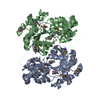
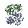
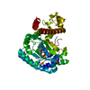
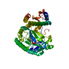
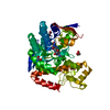
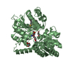
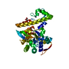
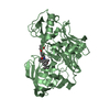
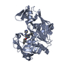
| Title | Crystal Structure of CalG3, Calicheamicin Glycostyltransferase, TDP and calicheamicin T0 bound form | ||||||
 Components Components | CalG3 | ||||||
 Keywords Keywords | transferase/antibiotic / Calicheamicin / TDP / Structural Genomics / PSI-2 / Protein Structure Initiative / Center for Eukaryotic Structural Genomics / CESG / GT-B fold / Glycosyltransferase / transferase-antibiotic complex / Enzyme Discovery for Natural Product Biosynthesis / NatPro | ||||||
| Function / homology |  Function and homology information Function and homology informationUDP-glycosyltransferase activity / glucosyltransferase activity / antibiotic biosynthetic process Similarity search - Function | ||||||
| Biological species |  Micromonospora echinospora (bacteria) Micromonospora echinospora (bacteria) | ||||||
| Method |  X-RAY DIFFRACTION / SYNCHROTRON / MOLECULAR REPLACEMENT / molecular replacement / Resolution: 1.597 Å X-RAY DIFFRACTION / SYNCHROTRON / MOLECULAR REPLACEMENT / molecular replacement / Resolution: 1.597 Å | ||||||
 Authors Authors | Chang, A. / Singh, S. / Bingman, C.A. / Thorson, J.S. / Phillips Jr., G.N. / Center for Eukaryotic Structural Genomics (CESG) / Enzyme Discovery for Natural Product Biosynthesis (NatPro) | ||||||
 Citation Citation | Journal: Proc.Natl.Acad.Sci.USA / Year: 2011 Title: Complete set of glycosyltransferase structures in the calicheamicin biosynthetic pathway reveals the origin of regiospecificity. Authors: Chang, A. / Singh, S. / Helmich, K.E. / Goff, R.D. / Bingman, C.A. / Thorson, J.S. / Phillips, G.N. | ||||||
| History |
|
- Structure visualization
| Structure viewer | Molecule: MolmilJmol/JSmol |
|---|
- Downloads & links
-Download
| PDBx/mmCIF format | 3oti.cif.gz | 184.8 KB | Display | PDBx/mmCIF format |
|---|---|---|---|---|
| PDB format | pdb3oti.ent.gz | 142.8 KB | Display | PDB format |
| PDBx/mmJSON format | 3oti.json.gz | Tree view | PDBx/mmJSON format | |
| Others |  Other downloads Other downloads |
-Validation report
| Arichive directory | https://data.pdbj.org/pub/pdb/validation_reports/ot/3otiftp://data.pdbj.org/pub/pdb/validation_reports/ot/3oti | HTTPS FTP |
|---|
-Related structure data
| Related structure data |  3ia7C  3iaaC  3otgC  3othC 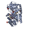 3rscC C: citing same article ( |
|---|---|
| Similar structure data | |
| Other databases |


-Links
 PDBj
PDBj- Assembly
| Deposited unit | 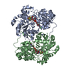
| ||||||||
|---|---|---|---|---|---|---|---|---|---|
| 1 | 
| ||||||||
| 2 | 
| ||||||||
| 3 |
| ||||||||
| Unit cell |
| ||||||||
| Details | The biological unit is a monomer. |
-Components
| #1: Protein | Mass: 43012.824 Da / Num. of mol.: 2 Source method: isolated from a genetically manipulated source Source: (gene. exp.) Micromonospora echinospora (bacteria) / Gene: calG3 / Plasmid: pET16b / Production host: #2: Chemical |   Mass: 402.188 Da / Num. of mol.: 2 / Source method: obtained synthetically / Formula: C10H16N2O11P2 Mass: 402.188 Da / Num. of mol.: 2 / Source method: obtained synthetically / Formula: C10H16N2O11P2#3: Chemical |   Mass: 584.682 Da / Num. of mol.: 2 / Source method: obtained synthetically / Formula: C24H28N2O9S3 Mass: 584.682 Da / Num. of mol.: 2 / Source method: obtained synthetically / Formula: C24H28N2O9S3#4: Chemical |   Mass: 35.453 Da / Num. of mol.: 2 / Source method: obtained synthetically / Formula: Cl Mass: 35.453 Da / Num. of mol.: 2 / Source method: obtained synthetically / Formula: Cl#5: Water | ChemComp-HOH / |  Mass: 18.015 Da / Num. of mol.: 941 / Source method: isolated from a natural source / Formula: H2O Mass: 18.015 Da / Num. of mol.: 941 / Source method: isolated from a natural source / Formula: H2O |
|---|
-Experimental details
-Experiment
| Experiment | Method: X-RAY DIFFRACTION / Number of used crystals: 1 |
|---|
- Sample preparation
| Crystal | Density Matthews: 2.21 Å3/Da / Density % sol: 44.35 % |
|---|---|
| Crystal grow | Temperature: 298 K / Method: vapor diffusion, hanging drop / pH: 4.5 Details: Protein Solution (10mg/ml CalG3 protein, 10mM Tris pH 7.5, 50mM NaCl, 25mM TDP, calicheamicin T0) mixed in a 1:1 ratio with the well solution (28% MEPEG2K, 160mM Na3Citrate, 100mM NaAcetate ...Details: Protein Solution (10mg/ml CalG3 protein, 10mM Tris pH 7.5, 50mM NaCl, 25mM TDP, calicheamicin T0) mixed in a 1:1 ratio with the well solution (28% MEPEG2K, 160mM Na3Citrate, 100mM NaAcetate pH 4.5) Cryoprotected with 20% Ethylene Glycol, 28% MEPEG2K, 160mM Na3Citrate, 100mM NaAcetate pH 4.5, vapor diffusion, hanging drop, temperature 298K, VAPOR DIFFUSION, HANGING DROP |
-Data collection
| Diffraction | Mean temperature: 100 K | |||||||||||||||||||||||||||||||||||||||||||||||||||||||||||||||||||||||||||||||||||||||||||||||||||||||||||||||||||||||||||||||||||||||||||||||||||
|---|---|---|---|---|---|---|---|---|---|---|---|---|---|---|---|---|---|---|---|---|---|---|---|---|---|---|---|---|---|---|---|---|---|---|---|---|---|---|---|---|---|---|---|---|---|---|---|---|---|---|---|---|---|---|---|---|---|---|---|---|---|---|---|---|---|---|---|---|---|---|---|---|---|---|---|---|---|---|---|---|---|---|---|---|---|---|---|---|---|---|---|---|---|---|---|---|---|---|---|---|---|---|---|---|---|---|---|---|---|---|---|---|---|---|---|---|---|---|---|---|---|---|---|---|---|---|---|---|---|---|---|---|---|---|---|---|---|---|---|---|---|---|---|---|---|---|---|---|
| Diffraction source | Source: SYNCHROTRON / Site: APS  / Beamline: 21-ID-D / Wavelength: 0.9794 Å / Beamline: 21-ID-D / Wavelength: 0.9794 Å | |||||||||||||||||||||||||||||||||||||||||||||||||||||||||||||||||||||||||||||||||||||||||||||||||||||||||||||||||||||||||||||||||||||||||||||||||||
| Detector | Type: MARMOSAIC 300 mm CCD / Detector: CCD / Date: Aug 20, 2010 / Details: mirrors and beryllium lenses | |||||||||||||||||||||||||||||||||||||||||||||||||||||||||||||||||||||||||||||||||||||||||||||||||||||||||||||||||||||||||||||||||||||||||||||||||||
| Radiation | Monochromator: C(111) / Protocol: SINGLE WAVELENGTH / Monochromatic (M) / Laue (L): M / Scattering type: x-ray | |||||||||||||||||||||||||||||||||||||||||||||||||||||||||||||||||||||||||||||||||||||||||||||||||||||||||||||||||||||||||||||||||||||||||||||||||||
| Radiation wavelength | Wavelength: 0.9794 Å / Relative weight: 1 | |||||||||||||||||||||||||||||||||||||||||||||||||||||||||||||||||||||||||||||||||||||||||||||||||||||||||||||||||||||||||||||||||||||||||||||||||||
| Reflection | Resolution: 1.597→50 Å / Num. obs: 96516 / % possible obs: 98 % / Redundancy: 7.1 % / Rmerge(I) obs: 0.095 / Χ2: 1.018 / Net I/σ(I): 10.8 | |||||||||||||||||||||||||||||||||||||||||||||||||||||||||||||||||||||||||||||||||||||||||||||||||||||||||||||||||||||||||||||||||||||||||||||||||||
| Reflection shell |
|
-Phasing
| Phasing | Method: molecular replacement | |||||||||
|---|---|---|---|---|---|---|---|---|---|---|
| Phasing MR |
|
- Processing
| Software |
| |||||||||||||||||||||||||||||||||||||||||||||||||||||||||||||||||||||||||||||||||||||||||||||||||||||||||
|---|---|---|---|---|---|---|---|---|---|---|---|---|---|---|---|---|---|---|---|---|---|---|---|---|---|---|---|---|---|---|---|---|---|---|---|---|---|---|---|---|---|---|---|---|---|---|---|---|---|---|---|---|---|---|---|---|---|---|---|---|---|---|---|---|---|---|---|---|---|---|---|---|---|---|---|---|---|---|---|---|---|---|---|---|---|---|---|---|---|---|---|---|---|---|---|---|---|---|---|---|---|---|---|---|---|---|
| Refinement | Method to determine structure: MOLECULAR REPLACEMENT / Resolution: 1.597→29.234 Å / Occupancy max: 1 / Occupancy min: 0.32 / SU ML: 0.17 / σ(F): 0.11 / Stereochemistry target values: ML
| |||||||||||||||||||||||||||||||||||||||||||||||||||||||||||||||||||||||||||||||||||||||||||||||||||||||||
| Solvent computation | Shrinkage radii: 0.9 Å / VDW probe radii: 1.11 Å / Solvent model: FLAT BULK SOLVENT MODEL / Bsol: 49.437 Å2 / ksol: 0.378 e/Å3 | |||||||||||||||||||||||||||||||||||||||||||||||||||||||||||||||||||||||||||||||||||||||||||||||||||||||||
| Displacement parameters | Biso max: 86.75 Å2 / Biso mean: 18.584 Å2 / Biso min: 5.35 Å2
| |||||||||||||||||||||||||||||||||||||||||||||||||||||||||||||||||||||||||||||||||||||||||||||||||||||||||
| Refinement step | Cycle: LAST / Resolution: 1.597→29.234 Å
| |||||||||||||||||||||||||||||||||||||||||||||||||||||||||||||||||||||||||||||||||||||||||||||||||||||||||
| Refine LS restraints |
| |||||||||||||||||||||||||||||||||||||||||||||||||||||||||||||||||||||||||||||||||||||||||||||||||||||||||
| LS refinement shell | Refine-ID: X-RAY DIFFRACTION / Total num. of bins used: 14
|
