 Movie
Movie Controller


+ Open data

- Basic information
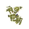
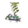
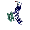
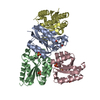
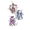
| Entry | Database: PDB / ID: 3onl | ||||||
|---|---|---|---|---|---|---|---|
| Title | yeast Ent3_ENTH-Vti1p_Habc complex structure | ||||||
 Components Components |
| ||||||
 Keywords Keywords | PROTEIN TRANSPORT / HELIX / ENTH / HABC / recognition between SNARE and adaptor | ||||||
| Function / homology |  Function and homology information Function and homology informationvacuolar calcium ion homeostasis / amphisome-lysosome fusion / Cargo recognition for clathrin-mediated endocytosis / Retrograde transport at the Trans-Golgi-Network / clathrin vesicle coat / Golgi vesicle fusion to target membrane / early endosome to Golgi transport / vesicle fusion with Golgi apparatus / vacuole fusion, non-autophagic / Platelet degranulation ...vacuolar calcium ion homeostasis / amphisome-lysosome fusion / Cargo recognition for clathrin-mediated endocytosis / Retrograde transport at the Trans-Golgi-Network / clathrin vesicle coat / Golgi vesicle fusion to target membrane / early endosome to Golgi transport / vesicle fusion with Golgi apparatus / vacuole fusion, non-autophagic / Platelet degranulation / Golgi to vacuole transport / late endosome to vacuole transport via multivesicular body sorting pathway / Golgi to endosome transport / multivesicular body sorting pathway / SNARE complex / SNAP receptor activity / vesicle fusion / phosphatidylinositol-3-phosphate binding / intra-Golgi vesicle-mediated transport / fungal-type vacuole membrane / phosphatidylinositol-3,5-bisphosphate binding / retrograde transport, endosome to Golgi / vacuolar membrane / clathrin binding / autophagosome membrane / SNARE binding / macroautophagy / ER to Golgi transport vesicle membrane / intracellular protein transport / phospholipid binding / endocytosis / late endosome membrane / protein transport / actin cytoskeleton organization / endosome / endosome membrane / Golgi membrane / endoplasmic reticulum membrane / Golgi apparatus / plasma membrane / cytosol Similarity search - Function | ||||||
| Biological species |  | ||||||
| Method |  X-RAY DIFFRACTION / SYNCHROTRON / MOLECULAR REPLACEMENT / Resolution: 2.2 Å X-RAY DIFFRACTION / SYNCHROTRON / MOLECULAR REPLACEMENT / Resolution: 2.2 Å | ||||||
 Authors Authors | Wang, J. / Fang, P. / Niu, L. / Teng, M. | ||||||
 Citation Citation | Journal: Proc.Natl.Acad.Sci.USA / Year: 2011 Title: Epsin N-terminal homology domains bind on opposite sides of two SNAREs Authors: Wang, J. / Gossing, M. / Fang, P. / Zimmermann, J. / Li, X. / von Mollard, G.F. / Niu, L. / Teng, M. | ||||||
| History |
|
- Structure visualization
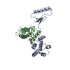
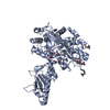
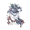
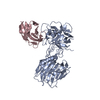
| Structure viewer | Molecule: MolmilJmol/JSmol |
|---|
- Downloads & links
-Download
| PDBx/mmCIF format | 3onl.cif.gz | 90.1 KB | Display | PDBx/mmCIF format |
|---|---|---|---|---|
| PDB format | pdb3onl.ent.gz | 68 KB | Display | PDB format |
| PDBx/mmJSON format | 3onl.json.gz | Tree view | PDBx/mmJSON format | |
| Others |  Other downloads Other downloads |
-Validation report
| Arichive directory | https://data.pdbj.org/pub/pdb/validation_reports/on/3onlftp://data.pdbj.org/pub/pdb/validation_reports/on/3onl | HTTPS FTP |
|---|
-Related structure data
| Related structure data |  3onjSC  3onkSC S: Starting model for refinement C: citing same article ( |
|---|---|
| Similar structure data |

-Links
 PDBj
PDBj


- Assembly
| Deposited unit | 
| ||||||||
|---|---|---|---|---|---|---|---|---|---|
| 1 |
| ||||||||
| Unit cell |
| ||||||||
| Details | THERE IS AN 1:1 COMPLEX FORMING BY TWO MONOMERIC PROTEIN. |
-Components
| #1: Protein | Mass: 17621.947 Da / Num. of mol.: 2 / Fragment: ENTH domain, residues 28-170 Source method: isolated from a genetically manipulated source Source: (gene. exp.) Plasmid: pET22b(+) / Production host:  #2: Protein | | Mass: 10995.161 Da / Num. of mol.: 1 / Fragment: Habc domain, residues 3-99 Source method: isolated from a genetically manipulated source Source: (gene. exp.) Plasmid: pET22b(+) / Production host: #3: Water | ChemComp-HOH / |  Mass: 18.015 Da / Num. of mol.: 180 / Source method: isolated from a natural source / Formula: H2O Mass: 18.015 Da / Num. of mol.: 180 / Source method: isolated from a natural source / Formula: H2O |
|---|
-Experimental details
-Experiment
| Experiment | Method: X-RAY DIFFRACTION / Number of used crystals: 1 |
|---|
- Sample preparation
| Crystal | Density Matthews: 2.51 Å3/Da / Density % sol: 50.94 % |
|---|---|
| Crystal grow | Temperature: 288 K / Method: vapor diffusion, hanging drop Details: 0.2M tri-Lithium Citrate tetrahydrate, 20% PEG 3350, VAPOR DIFFUSION, HANGING DROP, temperature 288.0K |
-Data collection
| Diffraction | Mean temperature: 100 K |
|---|---|
| Diffraction source | Source: SYNCHROTRON / Site: SSRF  / Beamline: BL17U / Wavelength: 1 Å / Beamline: BL17U / Wavelength: 1 Å |
| Detector | Type: MARMOSAIC 225 mm CCD / Detector: CCD / Date: Jan 15, 2010 |
| Radiation | Protocol: SINGLE WAVELENGTH / Monochromatic (M) / Laue (L): M / Scattering type: x-ray |
| Radiation wavelength | Wavelength: 1 Å / Relative weight: 1 |
| Reflection | Resolution: 2.2→50 Å / Num. obs: 23733 / % possible obs: 98.4 % |
| Reflection shell | Resolution: 2.2→2.28 Å / % possible all: 98.6 |
- Processing
| Software |
| |||||||||||||||||||||||||||||||||||||||||||||||||||||||||||||||||
|---|---|---|---|---|---|---|---|---|---|---|---|---|---|---|---|---|---|---|---|---|---|---|---|---|---|---|---|---|---|---|---|---|---|---|---|---|---|---|---|---|---|---|---|---|---|---|---|---|---|---|---|---|---|---|---|---|---|---|---|---|---|---|---|---|---|---|
| Refinement | Method to determine structure: MOLECULAR REPLACEMENT Starting model: PDB ENTRIES 3ONJ and 3ONK Resolution: 2.2→33.71 Å / Cor.coef. Fo:Fc: 0.931 / Cor.coef. Fo:Fc free: 0.903 / Occupancy max: 1 / Occupancy min: 0.59 / Cross valid method: THROUGHOUT / σ(F): 0 / ESU R Free: 0.228 / Stereochemistry target values: MAXIMUM LIKELIHOOD Details: HYDROGENS HAVE BEEN ADDED IN THE RIDING POSITIONS U VALUES: REFINED INDIVIDUALLY
| |||||||||||||||||||||||||||||||||||||||||||||||||||||||||||||||||
| Solvent computation | Ion probe radii: 0.8 Å / Shrinkage radii: 0.8 Å / VDW probe radii: 1.4 Å / Solvent model: MASK | |||||||||||||||||||||||||||||||||||||||||||||||||||||||||||||||||
| Displacement parameters | Biso max: 113.15 Å2 / Biso mean: 38.5679 Å2 / Biso min: 17.42 Å2
| |||||||||||||||||||||||||||||||||||||||||||||||||||||||||||||||||
| Refinement step | Cycle: LAST / Resolution: 2.2→33.71 Å
| |||||||||||||||||||||||||||||||||||||||||||||||||||||||||||||||||
| Refine LS restraints |
| |||||||||||||||||||||||||||||||||||||||||||||||||||||||||||||||||
| LS refinement shell | Resolution: 2.202→2.259 Å / Total num. of bins used: 20
|
