 Movie
Movie Controller

[English] 日本語
 Yorodumi
- PDB-3ol4: Crystal structure of a putative uncharacterized protein from Myco... -
+ Open data

- Basic information
| Entry | Database: PDB / ID: 3ol4 | ||||||
|---|---|---|---|---|---|---|---|
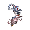
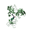
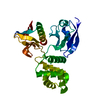
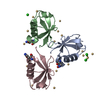
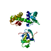
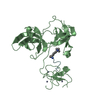
| Title | Crystal structure of a putative uncharacterized protein from Mycobacterium smegmatis, an ortholog of Rv0543c | ||||||
 Components Components | Putative uncharacterized protein | ||||||
 Keywords Keywords | UNKNOWN FUNCTION / mycobacterium tuberculosis / ortholog / Rv0543c / Structural Genomics / Seattle Structural Genomics Center for Infectious Disease / SSGCID | ||||||
| Function / homology |  Function and homology information Function and homology informationArc Repressor Mutant, subunit A - #2390 / Helix Hairpins - #2080 / Protein of unknown function DUF3349 / Protein of unknown function (DUF3349) / Helix Hairpins / Helix non-globular / Special / Arc Repressor Mutant, subunit A / Orthogonal Bundle / Mainly Alpha Similarity search - Domain/homology | ||||||
| Biological species |  Mycobacterium smegmatis (bacteria) Mycobacterium smegmatis (bacteria) | ||||||
| Method |  X-RAY DIFFRACTION / MOLECULAR REPLACEMENT / molecular replacement / Resolution: 2 Å X-RAY DIFFRACTION / MOLECULAR REPLACEMENT / molecular replacement / Resolution: 2 Å | ||||||
 Authors Authors | Seattle Structural Genomics Center for Infectious Disease (SSGCID) | ||||||
 Citation Citation | Journal: Protein Sci. / Year: 2020 Title: Structural diversity in the Mycobacteria DUF3349 superfamily. Authors: Buchko, G.W. / Abendroth, J. / Robinson, J.I. / Phan, I.Q. / Myler, P.J. / Edwards, T.E. #1: Journal: Tuberculosis (Edinb) / Year: 2015Title: Increasing the structural coverage of tuberculosis drug targets. Authors: Baugh, L. / Phan, I. / Begley, D.W. / Clifton, M.C. / Armour, B. / Dranow, D.M. / Taylor, B.M. / Muruthi, M.M. / Abendroth, J. / Fairman, J.W. / Fox, D. / Dieterich, S.H. / Staker, B.L. / ...Authors: Baugh, L. / Phan, I. / Begley, D.W. / Clifton, M.C. / Armour, B. / Dranow, D.M. / Taylor, B.M. / Muruthi, M.M. / Abendroth, J. / Fairman, J.W. / Fox, D. / Dieterich, S.H. / Staker, B.L. / Gardberg, A.S. / Choi, R. / Hewitt, S.N. / Napuli, A.J. / Myers, J. / Barrett, L.K. / Zhang, Y. / Ferrell, M. / Mundt, E. / Thompkins, K. / Tran, N. / Lyons-Abbott, S. / Abramov, A. / Sekar, A. / Serbzhinskiy, D. / Lorimer, D. / Buchko, G.W. / Stacy, R. / Stewart, L.J. / Edwards, T.E. / Van Voorhis, W.C. / Myler, P.J. | ||||||
| History |
|
- Structure visualization
| Structure viewer | Molecule: MolmilJmol/JSmol |
|---|
- Downloads & links
-Download
| PDBx/mmCIF format | 3ol4.cif.gz | 89.6 KB | Display | PDBx/mmCIF format |
|---|---|---|---|---|
| PDB format | pdb3ol4.ent.gz | 68.7 KB | Display | PDB format |
| PDBx/mmJSON format | 3ol4.json.gz | Tree view | PDBx/mmJSON format | |
| Others |  Other downloads Other downloads |
-Validation report
| Arichive directory | https://data.pdbj.org/pub/pdb/validation_reports/ol/3ol4ftp://data.pdbj.org/pub/pdb/validation_reports/ol/3ol4 | HTTPS FTP |
|---|
-Related structure data
| Related structure data | 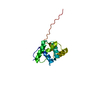 2lkyC  2m0nC  3ol3SC S: Starting model for refinement C: citing same article ( |
|---|---|
| Similar structure data | |
| Other databases |


-Links
 PDBj
PDBj- Assembly
| Deposited unit | 
| ||||||||
|---|---|---|---|---|---|---|---|---|---|
| 1 |
| ||||||||
| Unit cell |
| ||||||||
| Details | AUTHORS STATE THAT THE BIOLOGICAL MOLECULE IS UNKNOWN. |
-Components
| #1: Protein | Mass: 11695.176 Da / Num. of mol.: 2 Source method: isolated from a genetically manipulated source Source: (gene. exp.) Mycobacterium smegmatis (bacteria) / Strain: ATCC 700084 / mc(2)155 / Gene: MSMEG_1066 / Plasmid: AVA0421 / Production host: #2: Water | ChemComp-HOH / |  Mass: 18.015 Da / Num. of mol.: 169 / Source method: isolated from a natural source / Formula: H2O Mass: 18.015 Da / Num. of mol.: 169 / Source method: isolated from a natural source / Formula: H2O |
|---|
-Experimental details
-Experiment
| Experiment | Method: X-RAY DIFFRACTION / Number of used crystals: 1 |
|---|
- Sample preparation
| Crystal | Density Matthews: 2.5 Å3/Da / Density % sol: 50.74 % |
|---|---|
| Crystal grow | Temperature: 289 K / Method: vapor diffusion, sitting drop Details: 52.57 mg/mL MysmA,17112.a.A1 PS00688 against PACT screen condition F12, 0.2 M sodium malonate, 0.1 M BisTris Propane pH 6.5, 20% PEG 3350 with 25% ethylene glycol as cryo-protectant, crystal ...Details: 52.57 mg/mL MysmA,17112.a.A1 PS00688 against PACT screen condition F12, 0.2 M sodium malonate, 0.1 M BisTris Propane pH 6.5, 20% PEG 3350 with 25% ethylene glycol as cryo-protectant, crystal tracking ID 217010f12, VAPOR DIFFUSION, SITTING DROP, temperature 289K |
-Data collection
| Diffraction | Mean temperature: 100 K | ||||||||||||||||||||||||||||||||||||||||||||||||||||||||||||||||||||||||||||||||||||||||||||||||||||||||||||||||||||||||||||||||||||||||||||
|---|---|---|---|---|---|---|---|---|---|---|---|---|---|---|---|---|---|---|---|---|---|---|---|---|---|---|---|---|---|---|---|---|---|---|---|---|---|---|---|---|---|---|---|---|---|---|---|---|---|---|---|---|---|---|---|---|---|---|---|---|---|---|---|---|---|---|---|---|---|---|---|---|---|---|---|---|---|---|---|---|---|---|---|---|---|---|---|---|---|---|---|---|---|---|---|---|---|---|---|---|---|---|---|---|---|---|---|---|---|---|---|---|---|---|---|---|---|---|---|---|---|---|---|---|---|---|---|---|---|---|---|---|---|---|---|---|---|---|---|---|---|
| Diffraction source | Source: ROTATING ANODE / Type: RIGAKU FR-E+ SUPERBRIGHT / Wavelength: 1.5418 Å | ||||||||||||||||||||||||||||||||||||||||||||||||||||||||||||||||||||||||||||||||||||||||||||||||||||||||||||||||||||||||||||||||||||||||||||
| Detector | Type: RIGAKU SATURN 944+ / Detector: CCD / Date: Aug 11, 2010 / Details: VariMax HF | ||||||||||||||||||||||||||||||||||||||||||||||||||||||||||||||||||||||||||||||||||||||||||||||||||||||||||||||||||||||||||||||||||||||||||||
| Radiation | Protocol: SINGLE WAVELENGTH / Monochromatic (M) / Laue (L): M / Scattering type: x-ray | ||||||||||||||||||||||||||||||||||||||||||||||||||||||||||||||||||||||||||||||||||||||||||||||||||||||||||||||||||||||||||||||||||||||||||||
| Radiation wavelength | Wavelength: 1.5418 Å / Relative weight: 1 | ||||||||||||||||||||||||||||||||||||||||||||||||||||||||||||||||||||||||||||||||||||||||||||||||||||||||||||||||||||||||||||||||||||||||||||
| Reflection | Resolution: 2→50 Å / Num. all: 16421 / Num. obs: 16088 / % possible obs: 98 % / Observed criterion σ(I): -3 / Redundancy: 3.2 % / Biso Wilson estimate: 29.089 Å2 / Rmerge(I) obs: 0.048 / Net I/σ(I): 19.25 | ||||||||||||||||||||||||||||||||||||||||||||||||||||||||||||||||||||||||||||||||||||||||||||||||||||||||||||||||||||||||||||||||||||||||||||
| Reflection shell |
|
-Phasing
| Phasing | Method: molecular replacement |
|---|
- Processing
| Software |
| |||||||||||||||||||||||||||||||||||||||||||||||||||||||||||||||||||||||||||
|---|---|---|---|---|---|---|---|---|---|---|---|---|---|---|---|---|---|---|---|---|---|---|---|---|---|---|---|---|---|---|---|---|---|---|---|---|---|---|---|---|---|---|---|---|---|---|---|---|---|---|---|---|---|---|---|---|---|---|---|---|---|---|---|---|---|---|---|---|---|---|---|---|---|---|---|---|
| Refinement | Method to determine structure: MOLECULAR REPLACEMENT Starting model: PDB entry 3OL3 Resolution: 2→50 Å / Cor.coef. Fo:Fc: 0.94 / Cor.coef. Fo:Fc free: 0.922 / WRfactor Rfree: 0.2061 / WRfactor Rwork: 0.1766 / Occupancy max: 1 / Occupancy min: 0.3 / FOM work R set: 0.8283 / SU B: 9.434 / SU ML: 0.118 / SU R Cruickshank DPI: 0.1828 / SU Rfree: 0.1644 / Cross valid method: THROUGHOUT / σ(F): 0 / ESU R Free: 0.164 / Stereochemistry target values: MAXIMUM LIKELIHOOD Details: HYDROGENS HAVE BEEN ADDED IN THE RIDING POSITIONS. U VALUES WITH TLS ADDED.
| |||||||||||||||||||||||||||||||||||||||||||||||||||||||||||||||||||||||||||
| Solvent computation | Ion probe radii: 0.8 Å / Shrinkage radii: 0.8 Å / VDW probe radii: 1.4 Å / Solvent model: MASK | |||||||||||||||||||||||||||||||||||||||||||||||||||||||||||||||||||||||||||
| Displacement parameters | Biso max: 71.39 Å2 / Biso mean: 37.8828 Å2 / Biso min: 12.74 Å2
| |||||||||||||||||||||||||||||||||||||||||||||||||||||||||||||||||||||||||||
| Refinement step | Cycle: LAST / Resolution: 2→50 Å
| |||||||||||||||||||||||||||||||||||||||||||||||||||||||||||||||||||||||||||
| Refine LS restraints |
| |||||||||||||||||||||||||||||||||||||||||||||||||||||||||||||||||||||||||||
| LS refinement shell | Resolution: 2→2.052 Å / Total num. of bins used: 20
| |||||||||||||||||||||||||||||||||||||||||||||||||||||||||||||||||||||||||||
| Refinement TLS params. | Method: refined / Refine-ID: X-RAY DIFFRACTION
| |||||||||||||||||||||||||||||||||||||||||||||||||||||||||||||||||||||||||||
| Refinement TLS group |
|
