 Movie
Movie Controller

[English] 日本語
 Yorodumi
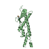
- PDB-3ogi: Crystal structure of the Mycobacterium tuberculosis H37Rv EsxOP c... -
+ Open data

- Basic information
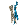
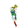
| Entry | Database: PDB / ID: 3ogi | ||||||
|---|---|---|---|---|---|---|---|
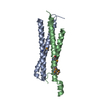
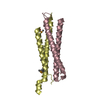
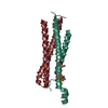
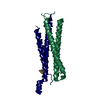
| Title | Crystal structure of the Mycobacterium tuberculosis H37Rv EsxOP complex (Rv2346c-Rv2347c) | ||||||
 Components Components |
| ||||||
 Keywords Keywords | Structural Genomics / Unknown function / PSI-2 / Protein Structure Initiative / Integrated Center for Structure and Function Innovation / ISFI / TB Structural Genomics Consortium / TBSGC / WXG100 / Secreted | ||||||
| Function / homology |  Function and homology information Function and homology informationcell wall / peptidoglycan-based cell wall / extracellular region / plasma membrane Similarity search - Function | ||||||
| Biological species |   Mycobacterium tuberculosis (bacteria) Mycobacterium tuberculosis (bacteria) | ||||||
| Method |  X-RAY DIFFRACTION / SYNCHROTRON / MAD / Resolution: 2.549 Å X-RAY DIFFRACTION / SYNCHROTRON / MAD / Resolution: 2.549 Å | ||||||
 Authors Authors | Arbing, M.A. / Chan, S. / Zhou, T.T. / Ahn, C. / Harris, L. / Kuo, E. / Sawaya, M.R. / Cascio, D. / Eisenberg, D. / Integrated Center for Structure and Function Innovation (ISFI) / TB Structural Genomics Consortium (TBSGC) | ||||||
 Citation Citation | Journal: Plos One / Year: 2013 Title: Heterologous expression of mycobacterial Esx complexes in Escherichia coli for structural studies is facilitated by the use of maltose binding protein fusions. Authors: Arbing, M.A. / Chan, S. / Harris, L. / Kuo, E. / Zhou, T.T. / Ahn, C.J. / Nguyen, L. / He, Q. / Lu, J. / Menchavez, P.T. / Shin, A. / Holton, T. / Sawaya, M.R. / Cascio, D. / Eisenberg, D. | ||||||
| History |
|
- Structure visualization
| Structure viewer | Molecule: MolmilJmol/JSmol |
|---|
- Downloads & links
-Download
| PDBx/mmCIF format | 3ogi.cif.gz | 121.9 KB | Display | PDBx/mmCIF format |
|---|---|---|---|---|
| PDB format | pdb3ogi.ent.gz | 96.5 KB | Display | PDB format |
| PDBx/mmJSON format | 3ogi.json.gz | Tree view | PDBx/mmJSON format | |
| Others |  Other downloads Other downloads |
-Validation report
| Arichive directory | https://data.pdbj.org/pub/pdb/validation_reports/og/3ogiftp://data.pdbj.org/pub/pdb/validation_reports/og/3ogi | HTTPS FTP |
|---|
-Related structure data
| Related structure data | 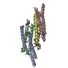 3q4hC 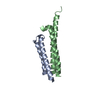 4gzrC 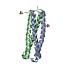 4i0xC C: citing same article ( |
|---|---|
| Similar structure data | |
| Other databases |

-Links
 PDBj
PDBj- Assembly
| Deposited unit | 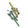
| ||||||||
|---|---|---|---|---|---|---|---|---|---|
| 1 | 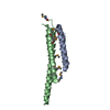
| ||||||||
| 2 | 
| ||||||||
| Unit cell |
|
-Components
| #1: Protein | Mass: 11031.625 Da / Num. of mol.: 2 Source method: isolated from a genetically manipulated source Source: (gene. exp.) Mycobacterium tuberculosis (bacteria) / Strain: H37Rv / Gene: MT2411, MTCY98.15c, Rv2346c / Plasmid: pMAPLe3 / Production host: #2: Protein | Mass: 11497.335 Da / Num. of mol.: 2 Source method: isolated from a genetically manipulated source Source: (gene. exp.) Mycobacterium tuberculosis (bacteria) / Strain: H37Rv / Gene: MT2412, MTCY98.16c, Rv2347c / Plasmid: pMAPLe3 / Production host: #3: Water | ChemComp-HOH / |  Mass: 18.015 Da / Num. of mol.: 35 / Source method: isolated from a natural source / Formula: H2O Mass: 18.015 Da / Num. of mol.: 35 / Source method: isolated from a natural source / Formula: H2OHas protein modification | Y | |
|---|
-Experimental details
-Experiment
| Experiment | Method: X-RAY DIFFRACTION / Number of used crystals: 1 |
|---|
- Sample preparation
| Crystal | Density Matthews: 2.3 Å3/Da / Density % sol: 46.42 % |
|---|---|
| Crystal grow | Temperature: 293 K / Method: vapor diffusion, hanging drop / pH: 4.6 Details: Reservoir: 9.0% isopropanol, 200 mM CaCl2, 90 mM sodium acetate trihydrate pH 4.6. Protein buffer: 50 mM HEPES pH 7.8, 150 mM NaCl. Ratio = 2:1 protein solution to reservoir solution, VAPOR ...Details: Reservoir: 9.0% isopropanol, 200 mM CaCl2, 90 mM sodium acetate trihydrate pH 4.6. Protein buffer: 50 mM HEPES pH 7.8, 150 mM NaCl. Ratio = 2:1 protein solution to reservoir solution, VAPOR DIFFUSION, HANGING DROP, temperature 293K |
-Data collection
| Diffraction | Mean temperature: 100 K | |||||||||
|---|---|---|---|---|---|---|---|---|---|---|
| Diffraction source | Source: SYNCHROTRON / Site: APS  / Beamline: 24-ID-C / Wavelength: 0.97918, 0.97938 / Beamline: 24-ID-C / Wavelength: 0.97918, 0.97938 | |||||||||
| Detector | Type: ADSC QUANTUM 315 / Detector: CCD / Date: Apr 26, 2010 | |||||||||
| Radiation | Monochromator: CRYO-COOLED SI(111) DOUBLE CRYSTAL / Protocol: MAD / Monochromatic (M) / Laue (L): M / Scattering type: x-ray | |||||||||
| Radiation wavelength |
| |||||||||
| Reflection | Resolution: 2.55→100 Å / Num. obs: 25291 / % possible obs: 100 % / Observed criterion σ(I): -3 / Redundancy: 5.5 % / Biso Wilson estimate: 55.54 Å2 / Rmerge(I) obs: 0.078 | |||||||||
| Reflection shell | Resolution: 2.55→2.64 Å / Rmerge(I) obs: 0.449 / Mean I/σ(I) obs: 3.4 / % possible all: 96.5 |
- Processing
| Software |
| |||||||||||||||||||||||||||||||||||||||||||||||||||||||||||||||||||||||||||||||||||||||||||||||||||||||||||||||||||||||||||||
|---|---|---|---|---|---|---|---|---|---|---|---|---|---|---|---|---|---|---|---|---|---|---|---|---|---|---|---|---|---|---|---|---|---|---|---|---|---|---|---|---|---|---|---|---|---|---|---|---|---|---|---|---|---|---|---|---|---|---|---|---|---|---|---|---|---|---|---|---|---|---|---|---|---|---|---|---|---|---|---|---|---|---|---|---|---|---|---|---|---|---|---|---|---|---|---|---|---|---|---|---|---|---|---|---|---|---|---|---|---|---|---|---|---|---|---|---|---|---|---|---|---|---|---|---|---|---|
| Refinement | Method to determine structure: MAD / Resolution: 2.549→28.307 Å / SU ML: 0.36 / σ(F): 0 / Phase error: 22.59 / Stereochemistry target values: ML
| |||||||||||||||||||||||||||||||||||||||||||||||||||||||||||||||||||||||||||||||||||||||||||||||||||||||||||||||||||||||||||||
| Solvent computation | Shrinkage radii: 0.9 Å / VDW probe radii: 1.11 Å / Solvent model: FLAT BULK SOLVENT MODEL / Bsol: 59.073 Å2 / ksol: 0.333 e/Å3 | |||||||||||||||||||||||||||||||||||||||||||||||||||||||||||||||||||||||||||||||||||||||||||||||||||||||||||||||||||||||||||||
| Displacement parameters |
| |||||||||||||||||||||||||||||||||||||||||||||||||||||||||||||||||||||||||||||||||||||||||||||||||||||||||||||||||||||||||||||
| Refinement step | Cycle: LAST / Resolution: 2.549→28.307 Å
| |||||||||||||||||||||||||||||||||||||||||||||||||||||||||||||||||||||||||||||||||||||||||||||||||||||||||||||||||||||||||||||
| Refine LS restraints |
| |||||||||||||||||||||||||||||||||||||||||||||||||||||||||||||||||||||||||||||||||||||||||||||||||||||||||||||||||||||||||||||
| LS refinement shell |
| |||||||||||||||||||||||||||||||||||||||||||||||||||||||||||||||||||||||||||||||||||||||||||||||||||||||||||||||||||||||||||||
| Refinement TLS params. | Method: refined / Refine-ID: X-RAY DIFFRACTION
| |||||||||||||||||||||||||||||||||||||||||||||||||||||||||||||||||||||||||||||||||||||||||||||||||||||||||||||||||||||||||||||
| Refinement TLS group |
|
