 Movie
Movie Controller

[English] 日本語
 Yorodumi
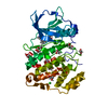
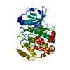
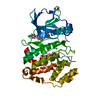
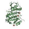
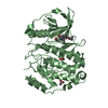
- PDB-3ofm: Structure of a human CK2alpha prime, the paralog isoform of the c... -
+ Open data

- Basic information
| Entry | Database: PDB / ID: 3ofm | ||||||
|---|---|---|---|---|---|---|---|
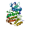
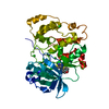
| Title | Structure of a human CK2alpha prime, the paralog isoform of the catalytic subunit of protein kinase CK2 from Homo sapiens | ||||||
 Components Components | Casein kinase II subunit alpha' | ||||||
 Keywords Keywords | transferase/transferase inhibitor / eukaryptic protein kinase fold / phospho transferase / ATP binding / Phosphorylation / Cytoplasm and nucleus / transferase-transferase inhibitor complex | ||||||
| Function / homology |  Function and homology information Function and homology informationregulation of mitophagy / regulation of chromosome separation / WNT mediated activation of DVL / Condensation of Prometaphase Chromosomes / protein kinase CK2 complex / : / Phosphorylation and nuclear translocation of the CRY:PER:kinase complex / Regulation of CDH1 posttranslational processing and trafficking to plasma membrane / Receptor Mediated Mitophagy / Synthesis of PC ...regulation of mitophagy / regulation of chromosome separation / WNT mediated activation of DVL / Condensation of Prometaphase Chromosomes / protein kinase CK2 complex / : / Phosphorylation and nuclear translocation of the CRY:PER:kinase complex / Regulation of CDH1 posttranslational processing and trafficking to plasma membrane / Receptor Mediated Mitophagy / Synthesis of PC / RUNX1 interacts with co-factors whose precise effect on RUNX1 targets is not known / Maturation of hRSV A proteins / negative regulation of apoptotic signaling pathway / negative regulation of proteasomal ubiquitin-dependent protein catabolic process / liver regeneration / acrosomal vesicle / Signal transduction by L1 / cerebral cortex development / Wnt signaling pathway / Regulation of PTEN stability and activity / KEAP1-NFE2L2 pathway / double-strand break repair / Cooperation of PDCL (PhLP1) and TRiC/CCT in G-protein beta folding / spermatogenesis / Regulation of TP53 Activity through Phosphorylation / non-specific serine/threonine protein kinase / protein serine kinase activity / protein serine/threonine kinase activity / apoptotic process / DNA damage response / chromatin / nucleoplasm / ATP binding / nucleus / cytosol Similarity search - Function | ||||||
| Biological species |  Homo sapiens (human) Homo sapiens (human) | ||||||
| Method |  X-RAY DIFFRACTION / SYNCHROTRON / MOLECULAR REPLACEMENT / Resolution: 2 Å X-RAY DIFFRACTION / SYNCHROTRON / MOLECULAR REPLACEMENT / Resolution: 2 Å | ||||||
 Authors Authors | Bischoff, N. / Olsen, B. / Raaf, J. / Bretner, M. / Issinger, O.-G. / Niefind, K. | ||||||
 Citation Citation | Journal: J.Mol.Biol. / Year: 2011 Title: Structural basis of the reduced affinity between the protein kinase CK2 subunits CK2alpha prime and CK2beta Authors: Bischoff, N. / Olsen, B. / Raaf, J. / Bretner, M. / Issinger, O.G. / Niefind, K. #1: Journal: Embo J. / Year: 2001Title: Crystal structure of human protein kinase CK2: insights into basic properties of the CK2 holoenzyme Authors: Niefind, K. / Guerra, B. / Ermakowa, I. / Issinger, O.-G. #2: Journal: Embo J. / Year: 1998 Title: Crystal structure of the catalytic subunit of protein kinase CK2 from Zea mays at 2.1 A resolution Authors: Niefind, K. / Guerra, B. / Pinna, L.A. / Issinger, O.-G. / Schomburg, D. #3: Journal: CELL.MOL.LIFE SCI. / Year: 2009 Title: Protein kinase CK2: from structures to insights Authors: Niefind, K. / Raaf, J. / Issinger, O.-G. | ||||||
| History |
|
- Structure visualization
| Structure viewer | Molecule: MolmilJmol/JSmol |
|---|
- Downloads & links
-Download
| PDBx/mmCIF format | 3ofm.cif.gz | 161.3 KB | Display | PDBx/mmCIF format |
|---|---|---|---|---|
| PDB format | pdb3ofm.ent.gz | 126.5 KB | Display | PDB format |
| PDBx/mmJSON format | 3ofm.json.gz | Tree view | PDBx/mmJSON format | |
| Others |  Other downloads Other downloads |
-Validation report
| Arichive directory | https://data.pdbj.org/pub/pdb/validation_reports/of/3ofmftp://data.pdbj.org/pub/pdb/validation_reports/of/3ofm | HTTPS FTP |
|---|
-Related structure data
| Related structure data |  2pvrS S: Starting model for refinement |
|---|---|
| Similar structure data |


-Links
 PDBj
PDBj


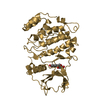
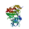
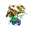
- Assembly
| Deposited unit | 
| ||||||||
|---|---|---|---|---|---|---|---|---|---|
| 1 |
| ||||||||
| Unit cell |
|
-Components
| #1: Protein | Mass: 41261.176 Da / Num. of mol.: 1 / Mutation: C336S Source method: isolated from a genetically manipulated source Source: (gene. exp.) Homo sapiens (human) / Gene: CK2A2, CSNK2A2 / Production host:  References: UniProt: P19784, non-specific serine/threonine protein kinase |
|---|---|
| #2: Chemical | ChemComp-4B0 / 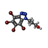  Mass: 492.787 Da / Num. of mol.: 1 / Source method: obtained synthetically / Formula: C9H7Br4N3O Mass: 492.787 Da / Num. of mol.: 1 / Source method: obtained synthetically / Formula: C9H7Br4N3O |
| #3: Chemical | ChemComp-CL /   Mass: 35.453 Da / Num. of mol.: 1 / Source method: obtained synthetically / Formula: Cl Mass: 35.453 Da / Num. of mol.: 1 / Source method: obtained synthetically / Formula: Cl |
| #4: Water | ChemComp-HOH /  Mass: 18.015 Da / Num. of mol.: 227 / Source method: isolated from a natural source / Formula: H2O Mass: 18.015 Da / Num. of mol.: 227 / Source method: isolated from a natural source / Formula: H2O |
-Experimental details
-Experiment
| Experiment | Method: X-RAY DIFFRACTION / Number of used crystals: 1 |
|---|
- Sample preparation
| Crystal | Density Matthews: 2.52 Å3/Da / Density % sol: 51.11 % |
|---|---|
| Crystal grow | Temperature: 293 K / Method: vapor diffusion, sitting drop / pH: 8.5 Details: Protein stock solution: 6 mg/ml protein, 25 mM Tris/HCl, pH 8.5, 500 mM sodium chloride, 1 mM inhibitor 3-(4,5,6,7-tetrabromo-1H-benzotriazol-1-yl)propan-1-ol Reservoir: 28 % PEG6000, 500 mM ...Details: Protein stock solution: 6 mg/ml protein, 25 mM Tris/HCl, pH 8.5, 500 mM sodium chloride, 1 mM inhibitor 3-(4,5,6,7-tetrabromo-1H-benzotriazol-1-yl)propan-1-ol Reservoir: 28 % PEG6000, 500 mM lithium chloride, 100 mM Tris/HCl, pH 8.5 Drop: mixture of 0.5 uL protein solution plus 0.5 uL reservoir, VAPOR DIFFUSION, SITTING DROP, temperature 293K |
-Data collection
| Diffraction | Mean temperature: 100 K |
|---|---|
| Diffraction source | Source: SYNCHROTRON / Site: BESSY  / Beamline: 14.1 / Wavelength: 0.91841 Å / Beamline: 14.1 / Wavelength: 0.91841 Å |
| Detector | Type: MARRESEARCH / Detector: CCD / Date: Jun 6, 2009 |
| Radiation | Monochromator: Si 111 CHANNEL / Protocol: SINGLE WAVELENGTH / Monochromatic (M) / Laue (L): M / Scattering type: x-ray |
| Radiation wavelength | Wavelength: 0.91841 Å / Relative weight: 1 |
| Reflection | Resolution: 2→33.03 Å / Num. all: 27265 / Num. obs: 23663 / % possible obs: 86.8 % / Observed criterion σ(I): -3 / Redundancy: 2.2 % / Biso Wilson estimate: 26.083 Å2 / Rmerge(I) obs: 0.057 / Rsym value: 0.057 / Net I/σ(I): 12.61 |
| Reflection shell | Resolution: 2→2.05 Å / Rmerge(I) obs: 0.257 / Mean I/σ(I) obs: 3.4 / % possible all: 47.7 |
- Processing
| Software |
| ||||||||||||||||||||||||||||||||||||||||||||||||||||||||||||||||||||||||||||||||||||||||||||||||||||||||||||||||||||||||||||||||||||||||||||||||||||||||||||||||||||||||||
|---|---|---|---|---|---|---|---|---|---|---|---|---|---|---|---|---|---|---|---|---|---|---|---|---|---|---|---|---|---|---|---|---|---|---|---|---|---|---|---|---|---|---|---|---|---|---|---|---|---|---|---|---|---|---|---|---|---|---|---|---|---|---|---|---|---|---|---|---|---|---|---|---|---|---|---|---|---|---|---|---|---|---|---|---|---|---|---|---|---|---|---|---|---|---|---|---|---|---|---|---|---|---|---|---|---|---|---|---|---|---|---|---|---|---|---|---|---|---|---|---|---|---|---|---|---|---|---|---|---|---|---|---|---|---|---|---|---|---|---|---|---|---|---|---|---|---|---|---|---|---|---|---|---|---|---|---|---|---|---|---|---|---|---|---|---|---|---|---|---|---|---|
| Refinement | Method to determine structure: MOLECULAR REPLACEMENT Starting model: PDB ENTRY 2PVR Resolution: 2→33.03 Å / Cor.coef. Fo:Fc: 0.947 / Cor.coef. Fo:Fc free: 0.929 / SU B: 7.974 / SU ML: 0.103 / Cross valid method: THROUGHOUT / σ(F): 0 / ESU R Free: 0.154 / Stereochemistry target values: MAXIMUM LIKELIHOOD / Details: HYDROGENS HAVE BEEN ADDED IN THE RIDING POSITIONS
| ||||||||||||||||||||||||||||||||||||||||||||||||||||||||||||||||||||||||||||||||||||||||||||||||||||||||||||||||||||||||||||||||||||||||||||||||||||||||||||||||||||||||||
| Solvent computation | Ion probe radii: 0.8 Å / Shrinkage radii: 0.8 Å / VDW probe radii: 1.4 Å / Solvent model: BABINET MODEL WITH MASK | ||||||||||||||||||||||||||||||||||||||||||||||||||||||||||||||||||||||||||||||||||||||||||||||||||||||||||||||||||||||||||||||||||||||||||||||||||||||||||||||||||||||||||
| Displacement parameters | Biso mean: 26.698 Å2
| ||||||||||||||||||||||||||||||||||||||||||||||||||||||||||||||||||||||||||||||||||||||||||||||||||||||||||||||||||||||||||||||||||||||||||||||||||||||||||||||||||||||||||
| Refinement step | Cycle: LAST / Resolution: 2→33.03 Å
| ||||||||||||||||||||||||||||||||||||||||||||||||||||||||||||||||||||||||||||||||||||||||||||||||||||||||||||||||||||||||||||||||||||||||||||||||||||||||||||||||||||||||||
| Refine LS restraints |
| ||||||||||||||||||||||||||||||||||||||||||||||||||||||||||||||||||||||||||||||||||||||||||||||||||||||||||||||||||||||||||||||||||||||||||||||||||||||||||||||||||||||||||
| LS refinement shell | Resolution: 2→2.052 Å / Total num. of bins used: 20 /
| ||||||||||||||||||||||||||||||||||||||||||||||||||||||||||||||||||||||||||||||||||||||||||||||||||||||||||||||||||||||||||||||||||||||||||||||||||||||||||||||||||||||||||
| Refinement TLS params. | Method: refined / Refine-ID: X-RAY DIFFRACTION
| ||||||||||||||||||||||||||||||||||||||||||||||||||||||||||||||||||||||||||||||||||||||||||||||||||||||||||||||||||||||||||||||||||||||||||||||||||||||||||||||||||||||||||
| Refinement TLS group |
|
