 Movie
Movie Controller

[English] 日本語
 Yorodumi
- PDB-3o8m: Crystal structure of monomeric KlHxk1 in crystal form XI with glu... -
+ Open data

- Basic information
| Entry | Database: PDB / ID: 3o8m | ||||||
|---|---|---|---|---|---|---|---|
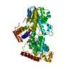
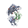
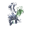
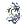
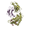
| Title | Crystal structure of monomeric KlHxk1 in crystal form XI with glucose bound (closed state) | ||||||
 Components Components | Hexokinase | ||||||
 Keywords Keywords | TRANSFERASE / RNASEH-LIKE FOLD / HEXOKINASE / GLYCOLYSIS / GLUCOSE REPRESSION / ATP BINDING / MIG1 BINDING | ||||||
| Function / homology |  Function and homology information Function and homology informationfructose import across plasma membrane / mannokinase activity / hexokinase / fructokinase activity / glucokinase activity / mannose metabolic process / D-glucose binding / fructose metabolic process / : / intracellular glucose homeostasis ...fructose import across plasma membrane / mannokinase activity / hexokinase / fructokinase activity / glucokinase activity / mannose metabolic process / D-glucose binding / fructose metabolic process / : / intracellular glucose homeostasis / glycolytic process / glucose metabolic process / mitochondrion / ATP binding / cytosol Similarity search - Function | ||||||
| Biological species |  Kluyveromyces lactis (yeast) Kluyveromyces lactis (yeast) | ||||||
| Method |  X-RAY DIFFRACTION / SYNCHROTRON / MOLECULAR REPLACEMENT / Resolution: 1.42 Å X-RAY DIFFRACTION / SYNCHROTRON / MOLECULAR REPLACEMENT / Resolution: 1.42 Å | ||||||
 Authors Authors | Kuettner, E.B. / Kettner, K. / Keim, A. / Kriegel, T.M. / Strater, N. | ||||||
 Citation Citation | Journal: J.Biol.Chem. / Year: 2010 Title: Crystal Structure of Hexokinase KlHxk1 of Kluyveromyces lactis: A MOLECULAR BASIS FOR UNDERSTANDING THE CONTROL OF YEAST HEXOKINASE FUNCTIONS VIA COVALENT MODIFICATION AND OLIGOMERIZATION. Authors: Kuettner, E.B. / Kettner, K. / Keim, A. / Svergun, D.I. / Volke, D. / Singer, D. / Hoffmann, R. / Muller, E.C. / Otto, A. / Kriegel, T.M. / Strater, N. #1: Journal: Acta Crystallogr.,Sect.F / Year: 2007 Title: Crystallization and preliminary X-ray diffraction studies of hexokinase KlHxk1 from Kluyveromyces lactis Authors: Kuettner, E.B. / Kriegel, T.M. / Keim, A. / Naumann, M. / Strater, N. | ||||||
| History |
|
- Structure visualization
| Structure viewer | Molecule: MolmilJmol/JSmol |
|---|
- Downloads & links
-Download
| PDBx/mmCIF format | 3o8m.cif.gz | 208.1 KB | Display | PDBx/mmCIF format |
|---|---|---|---|---|
| PDB format | pdb3o8m.ent.gz | 166.5 KB | Display | PDB format |
| PDBx/mmJSON format | 3o8m.json.gz | Tree view | PDBx/mmJSON format | |
| Others |  Other downloads Other downloads |
-Validation report
| Arichive directory | https://data.pdbj.org/pub/pdb/validation_reports/o8/3o8mftp://data.pdbj.org/pub/pdb/validation_reports/o8/3o8m | HTTPS FTP |
|---|
-Related structure data
| Related structure data |  3o08SC  3o1bC  3o1wC  3o4wC 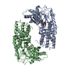 3o5bC 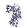 3o6wC 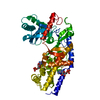 3o80C S: Starting model for refinement C: citing same article ( |
|---|---|
| Similar structure data |


-Links
 PDBj
PDBj


- Assembly
| Deposited unit | 
| ||||||||
|---|---|---|---|---|---|---|---|---|---|
| 1 |
| ||||||||
| Unit cell |
|
-Components
| #1: Protein | Mass: 53672.062 Da / Num. of mol.: 1 Source method: isolated from a genetically manipulated source Source: (gene. exp.) Kluyveromyces lactis (yeast) / Strain: CBS2359/152 / Gene: KLLA0D11352g, RAG5 / Plasmid: pTSRAG5 / Production host: Kluyveromyces lactis (yeast) / Strain (production host): JA6-delta-rag5 / References: UniProt: P33284, hexokinase | ||
|---|---|---|---|
| #2: Sugar | ChemComp-GLC /   Type: D-saccharide, alpha linking / Mass: 180.156 Da / Num. of mol.: 1 Type: D-saccharide, alpha linking / Mass: 180.156 Da / Num. of mol.: 1Source method: isolated from a genetically manipulated source Formula: C6H12O6 | ||
| #3: Sugar | ChemComp-BGC /   Type: D-saccharide, beta linking / Mass: 180.156 Da / Num. of mol.: 1 Type: D-saccharide, beta linking / Mass: 180.156 Da / Num. of mol.: 1Source method: isolated from a genetically manipulated source Formula: C6H12O6 | ||
| #4: Chemical | ChemComp-CL /   Mass: 35.453 Da / Num. of mol.: 4 / Source method: obtained synthetically / Formula: Cl Mass: 35.453 Da / Num. of mol.: 4 / Source method: obtained synthetically / Formula: Cl#5: Water | ChemComp-HOH / |  Mass: 18.015 Da / Num. of mol.: 611 / Source method: isolated from a natural source / Formula: H2O Mass: 18.015 Da / Num. of mol.: 611 / Source method: isolated from a natural source / Formula: H2O |
-Experimental details
-Experiment
| Experiment | Method: X-RAY DIFFRACTION / Number of used crystals: 1 |
|---|
- Sample preparation
| Crystal | Density Matthews: 2.36 Å3/Da / Density % sol: 47.92 % |
|---|---|
| Crystal grow | Temperature: 292 K / Method: vapor diffusion, hanging drop / pH: 7 Details: 0.2microL reservoir + 0.2microL protein, reservoir: 20% PEG6000, 1M LiCl, 0.1M Hepes pH 7.0, protein: 6.6mg/ml KlHxk1, 10mM Tris pH 7.4, 1mM EDTA, 1mM DTT, 0.5mM PMSF, 10mM AMPPNP, 10mM ...Details: 0.2microL reservoir + 0.2microL protein, reservoir: 20% PEG6000, 1M LiCl, 0.1M Hepes pH 7.0, protein: 6.6mg/ml KlHxk1, 10mM Tris pH 7.4, 1mM EDTA, 1mM DTT, 0.5mM PMSF, 10mM AMPPNP, 10mM Glucose, 10mM MgCl2, VAPOR DIFFUSION, HANGING DROP, temperature 292K |
-Data collection
| Diffraction | Mean temperature: 100 K |
|---|---|
| Diffraction source | Source: SYNCHROTRON / Site: BESSY  / Beamline: 14.2 / Wavelength: 0.91841 Å / Beamline: 14.2 / Wavelength: 0.91841 Å |
| Detector | Type: MARMOSAIC 225 mm CCD / Detector: CCD / Date: Jul 15, 2008 Details: 1st mirror: Silicon, active surface 50 nm Rh-coated, 2nd mirror: Glas, active surface 50 nm Rh-coated |
| Radiation | Monochromator: Si-111 crystal / Protocol: SINGLE WAVELENGTH / Monochromatic (M) / Laue (L): M / Scattering type: x-ray |
| Radiation wavelength | Wavelength: 0.91841 Å / Relative weight: 1 |
| Reflection | Resolution: 1.42→30 Å / Num. all: 96547 / Num. obs: 92838 / % possible obs: 96.2 % / Observed criterion σ(I): -3 / Redundancy: 7.1 % / Biso Wilson estimate: 23.1 Å2 / Rsym value: 0.05 / Net I/σ(I): 22.6 |
| Reflection shell | Resolution: 1.42→1.46 Å / Redundancy: 4.1 % / Mean I/σ(I) obs: 2.9 / Num. unique all: 5134 / Rsym value: 0.491 / % possible all: 72.7 |
- Processing
| Software |
| ||||||||||||||||||||||||||||||||||||||||||||||||||||||||||||||||||||||||||||||||||||||||||||||||||||||||||||||||||||||||||||||||||||||||||||||||||||||||||||||||||||||||||
|---|---|---|---|---|---|---|---|---|---|---|---|---|---|---|---|---|---|---|---|---|---|---|---|---|---|---|---|---|---|---|---|---|---|---|---|---|---|---|---|---|---|---|---|---|---|---|---|---|---|---|---|---|---|---|---|---|---|---|---|---|---|---|---|---|---|---|---|---|---|---|---|---|---|---|---|---|---|---|---|---|---|---|---|---|---|---|---|---|---|---|---|---|---|---|---|---|---|---|---|---|---|---|---|---|---|---|---|---|---|---|---|---|---|---|---|---|---|---|---|---|---|---|---|---|---|---|---|---|---|---|---|---|---|---|---|---|---|---|---|---|---|---|---|---|---|---|---|---|---|---|---|---|---|---|---|---|---|---|---|---|---|---|---|---|---|---|---|---|---|---|---|
| Refinement | Method to determine structure: MOLECULAR REPLACEMENT Starting model: PDB entry 3O08 Resolution: 1.42→29.7 Å / Cor.coef. Fo:Fc: 0.963 / Cor.coef. Fo:Fc free: 0.961 / SU B: 2.865 / SU ML: 0.051 / Cross valid method: THROUGHOUT / ESU R Free: 0.068 / Stereochemistry target values: MAXIMUM LIKELIHOOD Details: HYDROGENS HAVE BEEN ADDED IN THE RIDING POSITIONS. FULL B FACTORS AND ANISO RECORDS WERE COMPUTED BY CCP4 PROGRAMM TLSANL.
| ||||||||||||||||||||||||||||||||||||||||||||||||||||||||||||||||||||||||||||||||||||||||||||||||||||||||||||||||||||||||||||||||||||||||||||||||||||||||||||||||||||||||||
| Solvent computation | Ion probe radii: 0.8 Å / Shrinkage radii: 0.8 Å / VDW probe radii: 1.4 Å / Solvent model: MASK | ||||||||||||||||||||||||||||||||||||||||||||||||||||||||||||||||||||||||||||||||||||||||||||||||||||||||||||||||||||||||||||||||||||||||||||||||||||||||||||||||||||||||||
| Displacement parameters | Biso mean: 18.898 Å2
| ||||||||||||||||||||||||||||||||||||||||||||||||||||||||||||||||||||||||||||||||||||||||||||||||||||||||||||||||||||||||||||||||||||||||||||||||||||||||||||||||||||||||||
| Refinement step | Cycle: LAST / Resolution: 1.42→29.7 Å
| ||||||||||||||||||||||||||||||||||||||||||||||||||||||||||||||||||||||||||||||||||||||||||||||||||||||||||||||||||||||||||||||||||||||||||||||||||||||||||||||||||||||||||
| Refine LS restraints |
| ||||||||||||||||||||||||||||||||||||||||||||||||||||||||||||||||||||||||||||||||||||||||||||||||||||||||||||||||||||||||||||||||||||||||||||||||||||||||||||||||||||||||||
| LS refinement shell | Resolution: 1.42→1.457 Å / Total num. of bins used: 20
| ||||||||||||||||||||||||||||||||||||||||||||||||||||||||||||||||||||||||||||||||||||||||||||||||||||||||||||||||||||||||||||||||||||||||||||||||||||||||||||||||||||||||||
| Refinement TLS params. | Method: refined / Refine-ID: X-RAY DIFFRACTION
| ||||||||||||||||||||||||||||||||||||||||||||||||||||||||||||||||||||||||||||||||||||||||||||||||||||||||||||||||||||||||||||||||||||||||||||||||||||||||||||||||||||||||||
| Refinement TLS group |
|
