Type: MARMOSAIC 225 mm CCD / Detector: CCD / Date: Jan 24, 2010 / Details: double crystal monochromator
Radiation
Monochromator: double crystal monochromator / Protocol: SINGLE WAVELENGTH / Monochromatic (M) / Laue (L): M / Scattering type: x-ray
Radiation wavelength
Wavelength: 0.97949 Å / Relative weight: 1
Reflection
Resolution: 1.85→50 Å / Num. obs: 11409 / % possible obs: 99.9 % / Redundancy: 15.5 % / Rmerge(I) obs: 0.054 / Net I/σ(I): 16.7
Reflection shell
Resolution (Å)
Redundancy (%)
Rmerge(I) obs
Diffraction-ID
% possible all
1.85-1.89
14
0.496
1
99.9
1.89-1.94
15.6
0.398
1
100
1.94-1.99
15.8
0.314
1
100
1.99-2.05
16
0.23
1
100
2.05-2.12
15.9
0.181
1
100
2.12-2.19
16
0.136
1
100
2.19-2.28
16
0.111
1
100
2.28-2.39
15.9
0.095
1
100
2.39-2.51
15.9
0.074
1
100
2.51-2.67
15.9
0.065
1
100
2.67-2.87
15.8
0.062
1
100
2.87-3.16
15.7
0.067
1
100
3.16-3.62
15.5
0.06
1
100
3.62-4.56
15.2
0.036
1
100
4.56-50
13.6
0.035
1
99.1
-
Phasing
Phasing
Method: SAD
Phasing MAD
D res high: 1.85 Å / D res low: 29.16 Å / FOM acentric: 0.458 / FOM centric: 0.178 / Reflection acentric: 9304 / Reflection centric: 2013
Phasing MAD set
ID
R cullis acentric
R cullis centric
Highest resolution (Å)
Lowest resolution (Å)
Reflection acentric
Reflection centric
ISO_1
0
0
1.85
29.16
9304
2013
ANO_1
0.576
0
1.85
29.16
9304
0
Phasing MAD set shell
ID
Resolution (Å)
R cullis acentric
R cullis centric
Reflection acentric
Reflection centric
ISO_1
7.97-29.16
0
0
79
95
ISO_1
5.75-7.97
0
0
166
98
ISO_1
4.72-5.75
0
0
228
100
ISO_1
4.1-4.72
0
0
274
98
ISO_1
3.68-4.1
0
0
319
99
ISO_1
3.36-3.68
0
0
355
100
ISO_1
3.12-3.36
0
0
385
103
ISO_1
2.92-3.12
0
0
422
97
ISO_1
2.75-2.92
0
0
452
103
ISO_1
2.61-2.75
0
0
485
102
ISO_1
2.49-2.61
0
0
506
106
ISO_1
2.39-2.49
0
0
539
104
ISO_1
2.29-2.39
0
0
551
89
ISO_1
2.21-2.29
0
0
588
106
ISO_1
2.13-2.21
0
0
598
105
ISO_1
2.07-2.13
0
0
630
99
ISO_1
2.01-2.07
0
0
643
98
ISO_1
1.95-2.01
0
0
670
103
ISO_1
1.9-1.95
0
0
697
103
ISO_1
1.85-1.9
0
0
717
105
ANO_1
7.97-29.16
0.365
0
79
0
ANO_1
5.75-7.97
0.214
0
166
0
ANO_1
4.72-5.75
0.256
0
228
0
ANO_1
4.1-4.72
0.286
0
274
0
ANO_1
3.68-4.1
0.341
0
319
0
ANO_1
3.36-3.68
0.5
0
355
0
ANO_1
3.12-3.36
0.464
0
385
0
ANO_1
2.92-3.12
0.477
0
422
0
ANO_1
2.75-2.92
0.467
0
452
0
ANO_1
2.61-2.75
0.48
0
485
0
ANO_1
2.49-2.61
0.494
0
506
0
ANO_1
2.39-2.49
0.582
0
539
0
ANO_1
2.29-2.39
0.731
0
551
0
ANO_1
2.21-2.29
0.77
0
588
0
ANO_1
2.13-2.21
0.823
0
598
0
ANO_1
2.07-2.13
0.894
0
630
0
ANO_1
2.01-2.07
0.928
0
643
0
ANO_1
1.95-2.01
0.961
0
670
0
ANO_1
1.9-1.95
0.973
0
697
0
ANO_1
1.85-1.9
0.986
0
717
0
Phasing MAD set site
ID
Cartn x (Å)
Cartn y (Å)
Cartn z (Å)
Atom type symbol
B iso
Occupancy
1
40.823
54.637
9.021
ZN
37.2
2.44
2
41.456
61.059
-2.566
ZN
39.59
2.34
Phasing MAD shell
Resolution (Å)
FOM acentric
FOM centric
Reflection acentric
Reflection centric
7.97-29.16
0.725
0.224
79
95
5.75-7.97
0.745
0.183
166
98
4.72-5.75
0.655
0.22
228
100
4.1-4.72
0.659
0.176
274
98
3.68-4.1
0.61
0.151
319
99
3.36-3.68
0.552
0.153
355
100
3.12-3.36
0.567
0.152
385
103
2.92-3.12
0.572
0.183
422
97
2.75-2.92
0.594
0.194
452
103
2.61-2.75
0.598
0.22
485
102
2.49-2.61
0.601
0.187
506
106
2.39-2.49
0.564
0.188
539
104
2.29-2.39
0.471
0.168
551
89
2.21-2.29
0.449
0.187
588
106
2.13-2.21
0.41
0.165
598
105
2.07-2.13
0.354
0.175
630
99
2.01-2.07
0.318
0.147
643
98
1.95-2.01
0.279
0.155
670
103
1.9-1.95
0.254
0.167
697
103
1.85-1.9
0.236
0.163
717
105
Phasing dm
Method: Solvent flattening and Histogram matching / Reflection: 11317
Phasing dm shell
Resolution (Å)
Delta phi final
FOM
Reflection
5.48-100
58.6
0.798
501
4.28-5.48
56.4
0.91
513
3.71-4.28
55.1
0.91
508
3.35-3.71
55
0.92
509
3.1-3.35
56.6
0.913
514
2.91-3.1
49.5
0.904
504
2.76-2.91
54
0.908
501
2.64-2.76
53.4
0.892
502
2.53-2.64
54.5
0.886
508
2.44-2.53
48.6
0.871
508
2.36-2.44
55.5
0.833
508
2.29-2.36
61.2
0.833
501
2.23-2.29
59.2
0.849
501
2.17-2.23
57
0.853
508
2.12-2.17
60.8
0.866
512
2.07-2.12
61.4
0.855
508
2.03-2.07
67.6
0.854
517
1.99-2.03
65.5
0.851
516
1.95-1.99
66.4
0.831
516
1.92-1.95
66.7
0.817
540
1.88-1.92
73
0.78
550
1.85-1.88
76.4
0.708
572
-
Processing
Software
Name
Version
Classification
NB
DENZO
datareduction
SCALEPACK
datascaling
SHARP
phasing
DM
6.1
phasing
REFMAC
refmac_5.6.0081
refinement
PDB_EXTRACT
3.1
dataextraction
MxDC
datacollection
HKL-2000
datareduction
HKL-2000
datascaling
Refinement
Method to determine structure: SAD / Resolution: 1.85→27.38 Å / Cor.coef. Fo:Fc: 0.965 / Cor.coef. Fo:Fc free: 0.966 / Occupancy max: 1 / Occupancy min: 1 / SU B: 2.913 / SU ML: 0.053 / Cross valid method: THROUGHOUT / ESU R: 0.082 / ESU R Free: 0.076 / Stereochemistry target values: MAXIMUM LIKELIHOOD / Details: HYDROGENS HAVE BEEN USED IF PRESENT IN THE INPUT
Rfactor
Num. reflection
% reflection
Selection details
Rfree
0.18763
540
4.8 %
RANDOM
Rwork
0.18405
-
-
-
obs
0.18422
10775
99.96 %
-
Solvent computation
Ion probe radii: 0.8 Å / Shrinkage radii: 0.8 Å / VDW probe radii: 1.2 Å / Solvent model: BABINET MODEL WITH MASK
Displacement parameters
Biso mean: 43.436 Å2
Baniso -1
Baniso -2
Baniso -3
1-
0.01 Å2
0 Å2
0 Å2
2-
-
0.01 Å2
0 Å2
3-
-
-
-0.02 Å2
Refinement step
Cycle: LAST / Resolution: 1.85→27.38 Å
Protein
Nucleic acid
Ligand
Solvent
Total
Num. atoms
443
0
14
41
498
Refine LS restraints
Refine-ID
Type
Dev ideal
Dev ideal target
Number
X-RAY DIFFRACTION
r_bond_refined_d
0.011
0.022
464
X-RAY DIFFRACTION
r_bond_other_d
X-RAY DIFFRACTION
r_angle_refined_deg
1.215
1.965
620
X-RAY DIFFRACTION
r_angle_other_deg
X-RAY DIFFRACTION
r_dihedral_angle_1_deg
4.435
5
54
X-RAY DIFFRACTION
r_dihedral_angle_2_deg
32.148
22.632
19
X-RAY DIFFRACTION
r_dihedral_angle_3_deg
13.189
15
84
X-RAY DIFFRACTION
r_dihedral_angle_4_deg
13.227
15
3
X-RAY DIFFRACTION
r_chiral_restr
0.085
0.2
67
X-RAY DIFFRACTION
r_gen_planes_refined
0.005
0.021
337
X-RAY DIFFRACTION
r_gen_planes_other
X-RAY DIFFRACTION
r_nbd_refined
X-RAY DIFFRACTION
r_nbd_other
X-RAY DIFFRACTION
r_nbtor_refined
X-RAY DIFFRACTION
r_nbtor_other
X-RAY DIFFRACTION
r_xyhbond_nbd_refined
X-RAY DIFFRACTION
r_xyhbond_nbd_other
X-RAY DIFFRACTION
r_metal_ion_refined
X-RAY DIFFRACTION
r_metal_ion_other
X-RAY DIFFRACTION
r_symmetry_vdw_refined
X-RAY DIFFRACTION
r_symmetry_vdw_other
X-RAY DIFFRACTION
r_symmetry_hbond_refined
X-RAY DIFFRACTION
r_symmetry_hbond_other
X-RAY DIFFRACTION
r_symmetry_metal_ion_refined
X-RAY DIFFRACTION
r_symmetry_metal_ion_other
X-RAY DIFFRACTION
r_mcbond_it
X-RAY DIFFRACTION
r_mcbond_other
X-RAY DIFFRACTION
r_mcangle_it
X-RAY DIFFRACTION
r_scbond_it
X-RAY DIFFRACTION
r_scangle_it
X-RAY DIFFRACTION
r_rigid_bond_restr
X-RAY DIFFRACTION
r_sphericity_free
X-RAY DIFFRACTION
r_sphericity_bonded
LS refinement shell
Resolution: 1.85→1.898 Å / Total num. of bins used: 20
Rfactor
Num. reflection
% reflection
Rfree
0.289
33
-
Rwork
0.213
788
-
obs
-
-
99.88 %
Refinement TLS params.
Method: refined / Origin x: 3.696 Å / Origin y: 19.711 Å / Origin z: 34.077 Å
11
12
13
21
22
23
31
32
33
T
0.0948 Å2
-0.0131 Å2
0.0296 Å2
-
0.0337 Å2
-0.0199 Å2
-
-
0.0577 Å2
L
1.864 °2
-0.4929 °2
-0.5548 °2
-
1.1896 °2
0.3576 °2
-
-
3.1831 °2
S
0.0415 Å °
0.0492 Å °
-0.0618 Å °
-0.1132 Å °
-0.1485 Å °
0.1372 Å °
0.1948 Å °
-0.1676 Å °
0.107 Å °
+
About Yorodumi
-
News
-
Feb 9, 2022. New format data for meta-information of EMDB entries
New format data for meta-information of EMDB entries
Version 3 of the EMDB header file is now the official format.
The previous official version 1.9 will be removed from the archive.
In the structure databanks used in Yorodumi, some data are registered as the other names, "COVID-19 virus" and "2019-nCoV". Here are the details of the virus and the list of structure data.
Jan 31, 2019. EMDB accession codes are about to change! (news from PDBe EMDB page)
EMDB accession codes are about to change! (news from PDBe EMDB page)
The allocation of 4 digits for EMDB accession codes will soon come to an end. Whilst these codes will remain in use, new EMDB accession codes will include an additional digit and will expand incrementally as the available range of codes is exhausted. The current 4-digit format prefixed with “EMD-” (i.e. EMD-XXXX) will advance to a 5-digit format (i.e. EMD-XXXXX), and so on. It is currently estimated that the 4-digit codes will be depleted around Spring 2019, at which point the 5-digit format will come into force.
The EM Navigator/Yorodumi systems omit the EMD- prefix.
Related info.:Q: What is EMD? / ID/Accession-code notation in Yorodumi/EM Navigator
Yorodumi is a browser for structure data from EMDB, PDB, SASBDB, etc.
This page is also the successor to EM Navigator detail page, and also detail information page/front-end page for Omokage search.
The word "yorodu" (or yorozu) is an old Japanese word meaning "ten thousand". "mi" (miru) is to see.
Related info.:EMDB / PDB / SASBDB / Comparison of 3 databanks / Yorodumi Search / Aug 31, 2016. New EM Navigator & Yorodumi / Yorodumi Papers / Jmol/JSmol / Function and homology information / Changes in new EM Navigator and Yorodumi
 Movie
Movie Controller


 Open data
 Basic information
 Components
 Keywords
 Function and homology information
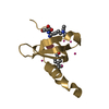
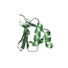
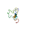
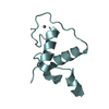
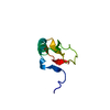
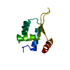
 Homo sapiens (human)
 X-RAY DIFFRACTION /
  Authors
 Citation
 Structure visualization
 Downloads & links
 Other downloads
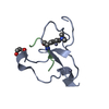


 PDBj
PDBj Assembly


 Mass: 92.094 Da / Num. of mol.: 2 / Source method: obtained synthetically / Formula: C3H8O3
 Mass: 65.409 Da / Num. of mol.: 2 / Source method: obtained synthetically / Formula: Zn
 Mass: 18.015 Da / Num. of mol.: 41 / Source method: isolated from a natural source / Formula: H2O
 Sample preparation
 / Beamline: 08ID-1 / Wavelength: 0.97949 Å
 Processing