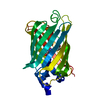
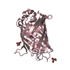
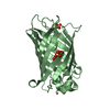
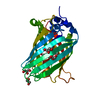
- PDB-3o59: DNA polymerase D large subunit DP2(1-300) from Pyrococcus horikoshii -
+
Open data
ID or keywords:
Loading...
-
Basic information
Entry
Database: PDB / ID: 3o59
Title
DNA polymerase D large subunit DP2(1-300) from Pyrococcus horikoshii
Components
DNA polymerase II large subunit
Keywords
TRANSFERASE / alpha helical structure / DNA POLYMERASE
Function / homology
Function and homology information
exodeoxyribonuclease I / intein-mediated protein splicing / single-stranded DNA 3'-5' DNA exonuclease activity / DNA catabolic process / DNA-templated DNA replication / DNA-directed DNA polymerase / DNA-directed DNA polymerase activity / DNA binding / identical protein binding Similarity search - Function
DNA polymerase II large subunit DP2 / DNA polymerase II large subunit DP2, N-terminal / : / : / DNA polymerase II large subunit DP2, N-terminal / DNA polymerase II large subunit DP2, central domain / DNA polymerase II large subunit DP2, catalytic domain / Intein C-terminal splicing region / Intein C-terminal splicing motif profile. / Hint domain C-terminal ...DNA polymerase II large subunit DP2 / DNA polymerase II large subunit DP2, N-terminal / : / : / DNA polymerase II large subunit DP2, N-terminal / DNA polymerase II large subunit DP2, central domain / DNA polymerase II large subunit DP2, catalytic domain / Intein C-terminal splicing region / Intein C-terminal splicing motif profile. / Hint domain C-terminal / Hint (Hedgehog/Intein) domain C-terminal region / Intein N-terminal splicing region / Intein N-terminal splicing motif profile. / Hint domain N-terminal / Hint (Hedgehog/Intein) domain N-terminal region / Hint domain superfamily / HNH nuclease Similarity search - Domain/homology
#1: Journal: J.Biol.Chem. / Year: 2003 Title: Subunit interaction and regulation of activity through terminal domains of the family D DNA polymerase from Pyrococcus horikoshii Authors: Shen, Y. / Tang, X.-F. / Matsui, I.
Mass: 18.015 Da / Num. of mol.: 107 / Source method: isolated from a natural source / Formula: H2O
Has protein modification
Y
Sequence details
ACCORDING TO THE GENBANK NP_142130, THE FIRST THREE RESIDUES, M-V-L, ARE INCLUDED AS THE ORIGINAL ...ACCORDING TO THE GENBANK NP_142130, THE FIRST THREE RESIDUES, M-V-L, ARE INCLUDED AS THE ORIGINAL SEQUENCE. THE FRAGMENT INFROMATION IS ADJUSTED FOR IT.
-
Experimental details
-
Experiment
Experiment
Method: X-RAY DIFFRACTION / Number of used crystals: 1
-
Sample preparation
Crystal
Density Matthews: 1.87 Å3/Da / Density % sol: 34.09 %
Crystal grow
Temperature: 293 K / Method: vapor diffusion, hanging drop / pH: 7.5 Details: 22.5% PEG 10000, 0.1M HEPES-NaOH, 40mM guanidine-HCl, pH 7.5, VAPOR DIFFUSION, HANGING DROP, temperature 293K
In the structure databanks used in Yorodumi, some data are registered as the other names, "COVID-19 virus" and "2019-nCoV". Here are the details of the virus and the list of structure data.
Jan 31, 2019. EMDB accession codes are about to change! (news from PDBe EMDB page)
EMDB accession codes are about to change! (news from PDBe EMDB page)
The allocation of 4 digits for EMDB accession codes will soon come to an end. Whilst these codes will remain in use, new EMDB accession codes will include an additional digit and will expand incrementally as the available range of codes is exhausted. The current 4-digit format prefixed with “EMD-” (i.e. EMD-XXXX) will advance to a 5-digit format (i.e. EMD-XXXXX), and so on. It is currently estimated that the 4-digit codes will be depleted around Spring 2019, at which point the 5-digit format will come into force.
The EM Navigator/Yorodumi systems omit the EMD- prefix.
Related info.:Q: What is EMD? / ID/Accession-code notation in Yorodumi/EM Navigator
Yorodumi is a browser for structure data from EMDB, PDB, SASBDB, etc.
This page is also the successor to EM Navigator detail page, and also detail information page/front-end page for Omokage search.
The word "yorodu" (or yorozu) is an old Japanese word meaning "ten thousand". "mi" (miru) is to see.
Related info.:EMDB / PDB / SASBDB / Comparison of 3 databanks / Yorodumi Search / Aug 31, 2016. New EM Navigator & Yorodumi / Yorodumi Papers / Jmol/JSmol / Function and homology information / Changes in new EM Navigator and Yorodumi
 Movie
Movie Controller

 Yorodumi
 Open data
 Basic information
 Components
 Keywords
 Function and homology information
 Pyrococcus horikoshii (archaea)
 X-RAY DIFFRACTION /
  Authors
 Citation
 Structure visualization
 Downloads & links
 Other downloads
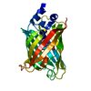


 PDBj
PDBj


 Assembly
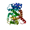

 Mass: 18.015 Da / Num. of mol.: 107 / Source method: isolated from a natural source / Formula: H2O
 Sample preparation
 / Beamline: BL-6A / Wavelength: 0.9788, 0.9793, 0.9900
 Processing