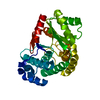
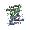
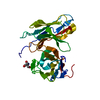
- PDB-3o0l: Crystal structure of a Pfam DUF1425 family member (Shew_1734) fro... -
+
Open data
ID or keywords:
Loading...
-
Basic information
Entry
Database: PDB / ID: 3o0l
Title
Crystal structure of a Pfam DUF1425 family member (Shew_1734) from Shewanella sp. pv-4 at 1.81 a resolution
Components
Uncharacterized protein
Keywords
STRUCTURAL GENOMICS / UNKNOWN FUNCTION / PFAM DUF1425 FAMILY MEMBER / JOINT CENTER FOR STRUCTURAL GENOMICS / JCSG / PROTEIN STRUCTURE INITIATIVE / PSI-BIOLOGY
Function / homology
Immunoglobulin-like - #3230 / Protein of unknown function DUF1425 / YcfL-like superfamily / Protein of unknown function (DUF1425) / Prokaryotic membrane lipoprotein lipid attachment site profile. / Immunoglobulin-like / Sandwich / Mainly Beta / DUF1425 domain-containing protein
Function and homology information
Biological species
Shewanella loihica (bacteria)
Method
X-RAY DIFFRACTION / SYNCHROTRON / MAD / Resolution: 1.81 Å
Mass: 18.015 Da / Num. of mol.: 157 / Source method: isolated from a natural source / Formula: H2O
Has protein modification
Y
Sequence details
THE CONSTRUCT (RESIDUES 26-335) WAS EXPRESSED WITH A PURIFICATION TAG MGSDKIHHHHHHENLYFQG. THE TAG ...THE CONSTRUCT (RESIDUES 26-335) WAS EXPRESSED WITH A PURIFICATION TAG MGSDKIHHHHHHENLYFQG. THE TAG WAS REMOVED WITH TEV PROTEASE LEAVING ONLY A GLYCINE (0) FOLLOWED BY THE TARGET SEQUENCE.
-
Experimental details
-
Experiment
Experiment
Method: X-RAY DIFFRACTION / Number of used crystals: 1
-
Sample preparation
Crystal
Density Matthews: 2.73 Å3/Da / Density % sol: 55.03 %
Type: MARMOSAIC 325 mm CCD / Detector: CCD / Date: Jun 11, 2010 / Details: Flat mirror (vertical focusing)
Radiation
Monochromator: Single crystal Si(111) bent monochromator (horizontal focusing) Protocol: MAD / Monochromatic (M) / Laue (L): M / Scattering type: x-ray
Radiation wavelength
ID
Wavelength (Å)
Relative weight
1
0.91837
1
2
0.9792
1
3
0.9786
1
Reflection
Resolution: 1.81→29.76 Å / Num. all: 25352 / Num. obs: 25352 / % possible obs: 99.9 % / Redundancy: 6 % / Biso Wilson estimate: 31.397 Å2 / Rsym value: 0.065 / Net I/σ(I): 13.1
Reflection shell
Diffraction-ID: 1
Resolution (Å)
Redundancy (%)
Rmerge(I) obs
Mean I/σ(I) obs
Num. measured all
Num. unique all
Rsym value
% possible all
1.81-1.86
6.1
0.74
2.2
11190
1835
0.74
100
1.86-1.91
6.1
0.755
1
11052
1811
0.755
100
1.91-1.96
6
0.421
1.7
10596
1754
0.421
100
1.96-2.02
6.1
0.32
2.4
10363
1700
0.32
100
2.02-2.09
6.1
0.253
3
10069
1656
0.253
100
2.09-2.16
6.1
0.187
3.9
9775
1596
0.187
100
2.16-2.25
6
0.188
3.7
9344
1545
0.188
100
2.25-2.34
6
0.172
3.4
8898
1485
0.172
100
2.34-2.44
6.1
0.114
6.2
8767
1435
0.114
100
2.44-2.56
6.1
0.093
7.3
8271
1355
0.093
100
2.56-2.7
6.1
0.084
7.8
7981
1314
0.084
100
2.7-2.86
6.1
0.077
8.2
7633
1260
0.077
100
2.86-3.06
6
0.071
8.7
6934
1150
0.071
100
3.06-3.3
6
0.058
10.7
6717
1111
0.058
100
3.3-3.62
6
0.05
11.7
6027
1005
0.05
100
3.62-4.05
6
0.043
14.2
5554
925
0.043
100
4.05-4.67
5.8
0.044
13.1
4823
825
0.044
100
4.67-5.72
5.8
0.045
13.1
4139
716
0.045
99.9
5.72-8.09
5.5
0.044
14.1
3094
560
0.044
100
8.09-29.76
4.7
0.047
14
1488
314
0.047
91.4
-
Phasing
Phasing
Method: MAD
-
Processing
Software
Name
Version
Classification
NB
SHELX
phasing
REFMAC
5.5.0110
refinement
SCALA
3.3.15
datascaling
PDB_EXTRACT
3.1
dataextraction
MOSFLM
datareduction
SHELXD
phasing
autoSHARP
phasing
Refinement
Method to determine structure: MAD / Resolution: 1.81→29.76 Å / Cor.coef. Fo:Fc: 0.961 / Cor.coef. Fo:Fc free: 0.955 / Occupancy max: 1 / Occupancy min: 0.25 / SU B: 6.01 / SU ML: 0.089 / Cross valid method: THROUGHOUT / σ(F): 0 / ESU R: 0.131 / ESU R Free: 0.12 / Phase error: 31.397 Stereochemistry target values: MAXIMUM LIKELIHOOD WITH PHASES Details: 1. HYDROGENS HAVE BEEN ADDED IN THE RIDING POSITIONS. 2. A MET-INHIBITION PROTOCOL WAS USED FOR SELENOMETHIONINE INCORPORATION DURING PROTEIN EXPRESSION. THE OCCUPANCY OF THE SE ATOMS IN THE ...Details: 1. HYDROGENS HAVE BEEN ADDED IN THE RIDING POSITIONS. 2. A MET-INHIBITION PROTOCOL WAS USED FOR SELENOMETHIONINE INCORPORATION DURING PROTEIN EXPRESSION. THE OCCUPANCY OF THE SE ATOMS IN THE MSE RESIDUES WAS REDUCED TO 0.75 FOR THE REDUCED SCATTERING POWER DUE TO PARTIAL S-MET INCORPORATION. 3. ATOM RECORD CONTAINS SUM OF TLS AND RESIDUAL B FACTORS. ANISOU RECORD CONTAINS SUM OF TLS AND RESIDUAL U FACTORS. 4. WATERS WERE EXCLUDED FROM AUTOMATIC TLS ASSIGNMENT. 5. ETHYLENE GLYCOL (EDO) MODELED IS PRESENT IN CRYSTALLIZATION/CRYO BUFFER.
Rfactor
Num. reflection
% reflection
Selection details
Rfree
0.2275
1276
5.1 %
RANDOM
Rwork
0.2041
-
-
-
obs
0.2052
25123
99.1 %
-
Solvent computation
Ion probe radii: 0.8 Å / Shrinkage radii: 0.8 Å / VDW probe radii: 1.2 Å / Solvent model: MASK
In the structure databanks used in Yorodumi, some data are registered as the other names, "COVID-19 virus" and "2019-nCoV". Here are the details of the virus and the list of structure data.
Jan 31, 2019. EMDB accession codes are about to change! (news from PDBe EMDB page)
EMDB accession codes are about to change! (news from PDBe EMDB page)
The allocation of 4 digits for EMDB accession codes will soon come to an end. Whilst these codes will remain in use, new EMDB accession codes will include an additional digit and will expand incrementally as the available range of codes is exhausted. The current 4-digit format prefixed with “EMD-” (i.e. EMD-XXXX) will advance to a 5-digit format (i.e. EMD-XXXXX), and so on. It is currently estimated that the 4-digit codes will be depleted around Spring 2019, at which point the 5-digit format will come into force.
The EM Navigator/Yorodumi systems omit the EMD- prefix.
Related info.:Q: What is EMD? / ID/Accession-code notation in Yorodumi/EM Navigator
Yorodumi is a browser for structure data from EMDB, PDB, SASBDB, etc.
This page is also the successor to EM Navigator detail page, and also detail information page/front-end page for Omokage search.
The word "yorodu" (or yorozu) is an old Japanese word meaning "ten thousand". "mi" (miru) is to see.
Related info.:EMDB / PDB / SASBDB / Comparison of 3 databanks / Yorodumi Search / Aug 31, 2016. New EM Navigator & Yorodumi / Yorodumi Papers / Jmol/JSmol / Function and homology information / Changes in new EM Navigator and Yorodumi
 Movie
Movie Controller

 Yorodumi
 Open data
 Basic information
 Components
 Keywords
 Function and homology information
 Shewanella loihica (bacteria)
 X-RAY DIFFRACTION /
  Authors
 Citation
 Structure visualization
 Downloads & links
 Other downloads
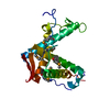


 PDBj
PDBj


 Assembly
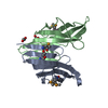

 Mass: 62.068 Da / Num. of mol.: 3 / Source method: obtained synthetically / Formula: C2H6O2
 Mass: 18.015 Da / Num. of mol.: 157 / Source method: isolated from a natural source / Formula: H2O
 Sample preparation
 / Beamline: BL11-1 / Wavelength: 0.91837,0.97920,0.97860
 Processing