electron transport chain / periplasmic space / electron transfer activity / iron ion binding / heme binding Similarity search - Function
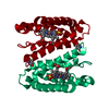
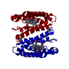
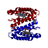
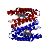
Cytochrome c prime, subgroup / Cytochrome c prime / Cytochrome c class II profile. / Cytochrome c, class II / Cytochrome C' / Cytochrome c/b562 / Cytochrome c/b562 / Four Helix Bundle (Hemerythrin (Met), subunit A) / Up-down Bundle / Mainly Alpha Similarity search - Domain/homology
Monochromator: SI MONOCHROMATOR / Protocol: SINGLE WAVELENGTH / Monochromatic (M) / Laue (L): M / Scattering type: x-ray
Radiation wavelength
Wavelength: 0.8 Å / Relative weight: 1
Reflection
Resolution: 1.22→45.1 Å / Num. obs: 47035 / % possible obs: 97.8 % / Redundancy: 4.5 % / Rmerge(I) obs: 0.03 / Net I/σ(I): 20
Reflection shell
Resolution: 1.22→1.29 Å / Redundancy: 3.3 % / Rmerge(I) obs: 0.51 / Mean I/σ(I) obs: 2.6 / % possible all: 88.4
-
Processing
Software
Name
Version
Classification
REFMAC
5.7.0032
refinement
XDS
datareduction
SCALA
datascaling
Refinement
Method to determine structure: OTHER Starting model: NONE Resolution: 1.22→45.09 Å / Cor.coef. Fo:Fc: 0.974 / Cor.coef. Fo:Fc free: 0.967 / SU B: 1.065 / SU ML: 0.021 / Cross valid method: THROUGHOUT / ESU R: 0.037 / ESU R Free: 0.037 / Stereochemistry target values: MAXIMUM LIKELIHOOD / Details: HYDROGENS HAVE BEEN ADDED IN THE RIDING POSITIONS.
Rfactor
Num. reflection
% reflection
Selection details
Rfree
0.17095
2309
5 %
RANDOM
Rwork
0.1441
-
-
-
obs
0.14541
43544
97.49 %
-
Solvent computation
Ion probe radii: 0.8 Å / Shrinkage radii: 0.8 Å / VDW probe radii: 1.2 Å / Solvent model: MASK
 Movie
Movie Controller

 Yorodumi
 Open data
 Basic information
 Components
 Keywords
 Function and homology information
 ACHROMOBACTER XYLOSOXIDANS (bacteria)
 X-RAY DIFFRACTION /
  Authors
 Citation
 Structure visualization
 Downloads & links
 Other downloads
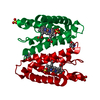


 PDBj
PDBj


 Assembly
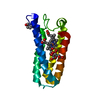


 Mass: 618.503 Da / Num. of mol.: 1 / Source method: obtained synthetically / Formula: C34H34FeN4O4
 Type: L-saccharide / Mass: 176.124 Da / Num. of mol.: 1 / Source method: obtained synthetically / Formula: C6H8O6
 Mass: 96.063 Da / Num. of mol.: 1 / Source method: obtained synthetically / Formula: SO4
 Mass: 18.015 Da / Num. of mol.: 166 / Source method: isolated from a natural source / Formula: H2O
 Sample preparation
 / Beamline: X10SA / Wavelength: 0.8
  Processing