 Movie
Movie Controller

[English] 日本語
 Yorodumi
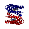
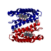
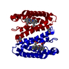
- PDB-4cdy: Spectroscopically-validated structure of cytochrome c prime from ... -
+ Open data

- Basic information
| Entry | Database: PDB / ID: 4cdy | |||||||||
|---|---|---|---|---|---|---|---|---|---|---|
| Title | Spectroscopically-validated structure of cytochrome c prime from Alcaligenes xylosoxidans, reduced by X-ray irradiation at 160K | |||||||||
 Components Components | CYTOCHROME C' | |||||||||
 Keywords Keywords | ELECTRON TRANSPORT / RESONANCE RAMAN / VALIDATION / GAS SENSOR / LIGAND DISCRIMINATION / FERRIC / HAEM / HEME / RADIOLYSIS | |||||||||
| Function / homology |  Function and homology information Function and homology informationelectron transport chain / electron transfer activity / periplasmic space / iron ion binding / heme binding Similarity search - Function | |||||||||
| Biological species |  ACHROMOBACTER XYLOSOXIDANS (bacteria) ACHROMOBACTER XYLOSOXIDANS (bacteria) | |||||||||
| Method |  X-RAY DIFFRACTION / SYNCHROTRON / MOLECULAR REPLACEMENT / Resolution: 1.77 Å X-RAY DIFFRACTION / SYNCHROTRON / MOLECULAR REPLACEMENT / Resolution: 1.77 Å | |||||||||
 Authors Authors | Kekilli, D. / Dworkowski, F. / Antonyuk, S. / Hough, M.A. | |||||||||
 Citation Citation | Journal: Acta Crystallogr.,Sect.D / Year: 2014 Title: Fingerprinting Redox and Ligand States in Haemprotein Crystal Structures Using Resonance Raman Spectroscopy. Authors: Kekilli, D. / Dworkowski, F.S. / Pompidor, G. / Fuchs, M.R. / Andrew, C.R. / Antonyuk, S. / Strange, R.W. / Eady, R.R. / Hasnain, S.S. / Hough, M.A. | |||||||||
| History |
|
- Structure visualization
| Structure viewer | Molecule: MolmilJmol/JSmol |
|---|
- Downloads & links
-Download
| PDBx/mmCIF format | 4cdy.cif.gz | 40.8 KB | Display | PDBx/mmCIF format |
|---|---|---|---|---|
| PDB format | pdb4cdy.ent.gz | 27.5 KB | Display | PDB format |
| PDBx/mmJSON format | 4cdy.json.gz | Tree view | PDBx/mmJSON format | |
| Others |  Other downloads Other downloads |
-Validation report
| Arichive directory | https://data.pdbj.org/pub/pdb/validation_reports/cd/4cdyftp://data.pdbj.org/pub/pdb/validation_reports/cd/4cdy | HTTPS FTP |
|---|
-Related structure data
| Related structure data | 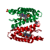 4cdaC 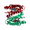 4cdvC 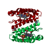 4cipC 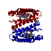 4cjgC 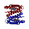 4cjoC 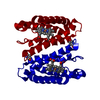 2yliS C: citing same article ( S: Starting model for refinement |
|---|---|
| Similar structure data |


-Links
 PDBj
PDBj


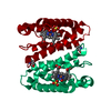
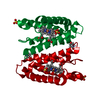
- Assembly
| Deposited unit | 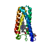
| ||||||||
|---|---|---|---|---|---|---|---|---|---|
| 1 | 
| ||||||||
| Unit cell |
|
-Components
| #1: Protein | Mass: 13631.442 Da / Num. of mol.: 1 Source method: isolated from a genetically manipulated source Source: (gene. exp.) ACHROMOBACTER XYLOSOXIDANS (bacteria) / Production host: |
|---|---|
| #2: Chemical | ChemComp-HEC /   Mass: 618.503 Da / Num. of mol.: 1 / Source method: obtained synthetically / Formula: C34H34FeN4O4 Mass: 618.503 Da / Num. of mol.: 1 / Source method: obtained synthetically / Formula: C34H34FeN4O4 |
| #3: Water | ChemComp-HOH /  Mass: 18.015 Da / Num. of mol.: 41 / Source method: isolated from a natural source / Formula: H2O Mass: 18.015 Da / Num. of mol.: 41 / Source method: isolated from a natural source / Formula: H2O |
| Has protein modification | Y |
-Experimental details
-Experiment
| Experiment | Method: X-RAY DIFFRACTION / Number of used crystals: 1 |
|---|
- Sample preparation
| Crystal | Density Matthews: 2.63 Å3/Da / Density % sol: 53.2 % / Description: NONE |
|---|---|
| Crystal grow | pH: 7.5 / Details: AMMONIUM SULFATE, HEPES PH 7.5 |
-Data collection
| Diffraction | Mean temperature: 160 K |
|---|---|
| Diffraction source | Source: SYNCHROTRON / Site: SLS  / Beamline: X10SA / Wavelength: 1 / Beamline: X10SA / Wavelength: 1 |
| Detector | Type: DECTRIS PILATUS 6M / Detector: PIXEL / Date: Mar 13, 2013 / Details: MIRRORS |
| Radiation | Monochromator: DOUBLE CRYSTAL / Protocol: SINGLE WAVELENGTH / Monochromatic (M) / Laue (L): M / Scattering type: x-ray |
| Radiation wavelength | Wavelength: 1 Å / Relative weight: 1 |
| Reflection | Resolution: 1.77→45 Å / Num. obs: 15687 / % possible obs: 98.6 % / Redundancy: 4.2 % / Rmerge(I) obs: 0.05 / Net I/σ(I): 11.7 |
| Reflection shell | Resolution: 1.77→1.87 Å / Redundancy: 4.4 % / Rmerge(I) obs: 0.7 / Mean I/σ(I) obs: 2 / % possible all: 79.4 |
- Processing
| Software |
| ||||||||||||||||||||||||||||||||||||||||||||||||||||||||||||||||||||||||||||||||||||||||||||||||||||||||||||||||||||||||||||||||||||||||||||||||||||||||||||||||||||||||||||||||||||||
|---|---|---|---|---|---|---|---|---|---|---|---|---|---|---|---|---|---|---|---|---|---|---|---|---|---|---|---|---|---|---|---|---|---|---|---|---|---|---|---|---|---|---|---|---|---|---|---|---|---|---|---|---|---|---|---|---|---|---|---|---|---|---|---|---|---|---|---|---|---|---|---|---|---|---|---|---|---|---|---|---|---|---|---|---|---|---|---|---|---|---|---|---|---|---|---|---|---|---|---|---|---|---|---|---|---|---|---|---|---|---|---|---|---|---|---|---|---|---|---|---|---|---|---|---|---|---|---|---|---|---|---|---|---|---|---|---|---|---|---|---|---|---|---|---|---|---|---|---|---|---|---|---|---|---|---|---|---|---|---|---|---|---|---|---|---|---|---|---|---|---|---|---|---|---|---|---|---|---|---|---|---|---|---|
| Refinement | Method to determine structure: MOLECULAR REPLACEMENT Starting model: PDB ENTRY 2YLI Resolution: 1.77→45.03 Å / Cor.coef. Fo:Fc: 0.956 / Cor.coef. Fo:Fc free: 0.941 / SU B: 2.521 / SU ML: 0.08 / Cross valid method: THROUGHOUT / ESU R: 0.116 / ESU R Free: 0.115 / Stereochemistry target values: MAXIMUM LIKELIHOOD Details: HYDROGENS HAVE BEEN ADDED IN THE RIDING POSITIONS. U VALUES REFINED INDIVIDUALLY
| ||||||||||||||||||||||||||||||||||||||||||||||||||||||||||||||||||||||||||||||||||||||||||||||||||||||||||||||||||||||||||||||||||||||||||||||||||||||||||||||||||||||||||||||||||||||
| Solvent computation | Ion probe radii: 0.8 Å / Shrinkage radii: 0.8 Å / VDW probe radii: 1.2 Å / Solvent model: MASK | ||||||||||||||||||||||||||||||||||||||||||||||||||||||||||||||||||||||||||||||||||||||||||||||||||||||||||||||||||||||||||||||||||||||||||||||||||||||||||||||||||||||||||||||||||||||
| Displacement parameters | Biso mean: 34.666 Å2
| ||||||||||||||||||||||||||||||||||||||||||||||||||||||||||||||||||||||||||||||||||||||||||||||||||||||||||||||||||||||||||||||||||||||||||||||||||||||||||||||||||||||||||||||||||||||
| Refinement step | Cycle: LAST / Resolution: 1.77→45.03 Å
| ||||||||||||||||||||||||||||||||||||||||||||||||||||||||||||||||||||||||||||||||||||||||||||||||||||||||||||||||||||||||||||||||||||||||||||||||||||||||||||||||||||||||||||||||||||||
| Refine LS restraints |
|