 Movie
Movie Controller

[English] 日本語
 Yorodumi
- PDB-3nys: X-ray structure of the K185A mutant of WbpE (WlbE) from pseudomon... -
+ Open data

- Basic information
| Entry | Database: PDB / ID: 3nys | ||||||
|---|---|---|---|---|---|---|---|
| Title | X-ray structure of the K185A mutant of WbpE (WlbE) from pseudomonas aeruginosa in complex with PLP at 1.45 angstrom resolution | ||||||
 Components Components | Aminotransferase WbpE | ||||||
 Keywords Keywords | TRANSFERASE / aminotransferase / PLP binding / nucleotide-sugar binding | ||||||
| Function / homology |  Function and homology information Function and homology informationUDP-2-acetamido-2-deoxy-ribo-hexuluronate aminotransferase / UDP-glucuronate biosynthetic process / O antigen biosynthetic process / lipopolysaccharide biosynthetic process / polysaccharide biosynthetic process / transaminase activity / cell wall organization / pyridoxal phosphate binding Similarity search - Function | ||||||
| Biological species |   Pseudomonas aeruginosa (bacteria) Pseudomonas aeruginosa (bacteria) | ||||||
| Method |  X-RAY DIFFRACTION / MOLECULAR REPLACEMENT / Resolution: 1.45 Å X-RAY DIFFRACTION / MOLECULAR REPLACEMENT / Resolution: 1.45 Å | ||||||
 Authors Authors | Holden, H.M. / Thoden, J.B. | ||||||
 Citation Citation | Journal: Protein Sci. / Year: 2017 Title: Structural investigation on WlaRG from Campylobacter jejuni: A sugar aminotransferase. Authors: Dow, G.T. / Gilbert, M. / Thoden, J.B. / Holden, H.M. | ||||||
| History |
|
- Structure visualization
| Structure viewer | Molecule: MolmilJmol/JSmol |
|---|
- Downloads & links
-Download
| PDBx/mmCIF format | 3nys.cif.gz | 94.7 KB | Display | PDBx/mmCIF format |
|---|---|---|---|---|
| PDB format | pdb3nys.ent.gz | 70 KB | Display | PDB format |
| PDBx/mmJSON format | 3nys.json.gz | Tree view | PDBx/mmJSON format | |
| Others |  Other downloads Other downloads |
-Validation report
| Arichive directory | https://data.pdbj.org/pub/pdb/validation_reports/ny/3nysftp://data.pdbj.org/pub/pdb/validation_reports/ny/3nys | HTTPS FTP |
|---|
-Related structure data
| Related structure data |  3nytC  3nyuC  5u1zC  5u20C  5u21C  5u23C  5u24C  3frkS C: citing same article ( S: Starting model for refinement |
|---|---|
| Similar structure data |


-Links
 PDBj
PDBj- Assembly
| Deposited unit | 
| |||||||||||||||
|---|---|---|---|---|---|---|---|---|---|---|---|---|---|---|---|---|
| 1 | 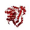
| |||||||||||||||
| Unit cell |
| |||||||||||||||
| Components on special symmetry positions |
|
-Components
| #1: Protein | Mass: 39978.586 Da / Num. of mol.: 1 / Mutation: K185A Source method: isolated from a genetically manipulated source Source: (gene. exp.) Pseudomonas aeruginosa (bacteria) / Strain: PAO1 / Gene: PA3155, wbpE / Plasmid: pET31 / Production host: |
|---|---|
| #2: Chemical | ChemComp-PLP /   Mass: 247.142 Da / Num. of mol.: 1 / Source method: obtained synthetically / Formula: C8H10NO6P Mass: 247.142 Da / Num. of mol.: 1 / Source method: obtained synthetically / Formula: C8H10NO6P |
| #3: Chemical | ChemComp-NA /   Mass: 22.990 Da / Num. of mol.: 1 / Source method: obtained synthetically / Formula: Na Mass: 22.990 Da / Num. of mol.: 1 / Source method: obtained synthetically / Formula: Na |
| #4: Water | ChemComp-HOH /  Mass: 18.015 Da / Num. of mol.: 485 / Source method: isolated from a natural source / Formula: H2O Mass: 18.015 Da / Num. of mol.: 485 / Source method: isolated from a natural source / Formula: H2O |
-Experimental details
-Experiment
| Experiment | Method: X-RAY DIFFRACTION / Number of used crystals: 1 |
|---|
- Sample preparation
| Crystal | Density Matthews: 2.31 Å3/Da / Density % sol: 46.74 % |
|---|---|
| Crystal grow | Temperature: 298 K / Method: vapor diffusion, hanging drop / pH: 6 Details: 25% pentaerythritrol propoxylate, 100 mM MES, pH 6, VAPOR DIFFUSION, HANGING DROP, temperature 298K |
-Data collection
| Diffraction | Mean temperature: 100 K |
|---|---|
| Diffraction source | Source: ROTATING ANODE / Type: RIGAKU RU200 / Wavelength: 1.54178 Å |
| Detector | Type: Bruker Platinum 135 / Detector: CCD / Date: Feb 22, 2010 / Details: mondel |
| Radiation | Protocol: SINGLE WAVELENGTH / Monochromatic (M) / Laue (L): M / Scattering type: x-ray |
| Radiation wavelength | Wavelength: 1.54178 Å / Relative weight: 1 |
| Reflection | Resolution: 1.45→30 Å / Num. all: 62362 / Num. obs: 62362 / % possible obs: 97.2 % / Observed criterion σ(F): 0 / Observed criterion σ(I): 0 / Redundancy: 5 % / Rmerge(I) obs: 0.061 / Rsym value: 0.061 / Net I/σ(I): 12.9 |
| Reflection shell | Resolution: 1.45→1.48 Å / Redundancy: 2.2 % / Rmerge(I) obs: 0.32 / Mean I/σ(I) obs: 2.7 / Num. unique all: 2316 / Rsym value: 0.32 / % possible all: 92.9 |
- Processing
| Software |
| |||||||||||||||||||||||||||||||||||||||||||||||||||||||||||||||||
|---|---|---|---|---|---|---|---|---|---|---|---|---|---|---|---|---|---|---|---|---|---|---|---|---|---|---|---|---|---|---|---|---|---|---|---|---|---|---|---|---|---|---|---|---|---|---|---|---|---|---|---|---|---|---|---|---|---|---|---|---|---|---|---|---|---|---|
| Refinement | Method to determine structure: MOLECULAR REPLACEMENT Starting model: PDB ENTRY 3FRK Resolution: 1.45→30 Å / Cor.coef. Fo:Fc: 0.958 / Cor.coef. Fo:Fc free: 0.94 / SU B: 1.148 / SU ML: 0.044 / Cross valid method: THROUGHOUT / σ(I): 0 / ESU R: 0.067 / ESU R Free: 0.07 / Stereochemistry target values: MAXIMUM LIKELIHOOD / Details: HYDROGENS HAVE BEEN ADDED IN THE RIDING POSITIONS
| |||||||||||||||||||||||||||||||||||||||||||||||||||||||||||||||||
| Solvent computation | Ion probe radii: 0.8 Å / Shrinkage radii: 0.8 Å / VDW probe radii: 1.4 Å / Solvent model: MASK | |||||||||||||||||||||||||||||||||||||||||||||||||||||||||||||||||
| Displacement parameters | Biso mean: 14.169 Å2
| |||||||||||||||||||||||||||||||||||||||||||||||||||||||||||||||||
| Refinement step | Cycle: LAST / Resolution: 1.45→30 Å
| |||||||||||||||||||||||||||||||||||||||||||||||||||||||||||||||||
| Refine LS restraints |
| |||||||||||||||||||||||||||||||||||||||||||||||||||||||||||||||||
| LS refinement shell | Resolution: 1.45→1.488 Å / Total num. of bins used: 20
|
