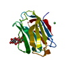
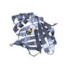
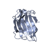
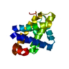
- PDB-3nv1: Crystal structure of human galectin-9 C-terminal CRD -
+
Open data
ID or keywords:
Loading...
-
Basic information
Entry
Database: PDB / ID: 3nv1
Title
Crystal structure of human galectin-9 C-terminal CRD
Components
Galectin 9 short isoform variant
Keywords
SUGAR BINDING PROTEIN / Sugar Binding
Function / homology
Function and homology information
positive regulation of activated T cell autonomous cell death / : / Interleukin-2 family signaling / positive regulation of dendritic cell apoptotic process / positive regulation of T cell activation via T cell receptor contact with antigen bound to MHC molecule on antigen presenting cell / negative regulation of mast cell degranulation / natural killer cell tolerance induction / negative regulation of T-helper 1 type immune response / NK T cell activation / positive regulation of dendritic cell differentiation ...positive regulation of activated T cell autonomous cell death / : / Interleukin-2 family signaling / positive regulation of dendritic cell apoptotic process / positive regulation of T cell activation via T cell receptor contact with antigen bound to MHC molecule on antigen presenting cell / negative regulation of mast cell degranulation / natural killer cell tolerance induction / negative regulation of T-helper 1 type immune response / NK T cell activation / positive regulation of dendritic cell differentiation / positive regulation of dendritic cell chemotaxis / galactoside binding / positive regulation of transforming growth factor beta production / galactose binding / oligosaccharide binding / negative regulation of chemokine production / negative regulation of CD4-positive, alpha-beta T cell proliferation / disaccharide binding / positive regulation of interleukin-13 production / positive regulation of CD4-positive, alpha-beta T cell proliferation / negative regulation of natural killer cell mediated cytotoxicity / positive regulation of monocyte chemotactic protein-1 production / negative regulation of activated T cell proliferation / positive regulation of interleukin-4 production / p38MAPK cascade / negative regulation of type II interferon production / positive regulation of interleukin-10 production / negative regulation of tumor necrosis factor production / maternal process involved in female pregnancy / ERK1 and ERK2 cascade / positive regulation of T cell migration / positive regulation of interleukin-12 production / : / response to interleukin-1 / positive regulation of interleukin-8 production / positive regulation of interleukin-1 beta production / female pregnancy / cellular response to virus / positive regulation of non-canonical NF-kappaB signal transduction / cellular response to type II interferon / positive regulation of interleukin-6 production / chemotaxis / positive regulation of type II interferon production / positive regulation of tumor necrosis factor production / carbohydrate binding / extracellular matrix / response to lipopolysaccharide / positive regulation of viral entry into host cell / positive regulation of ERK1 and ERK2 cascade / positive regulation of canonical NF-kappaB signal transduction / inflammatory response / receptor ligand activity / negative regulation of gene expression / positive regulation of gene expression / enzyme binding / : / nucleus / cytosol / cytoplasm Similarity search - Function
In the structure databanks used in Yorodumi, some data are registered as the other names, "COVID-19 virus" and "2019-nCoV". Here are the details of the virus and the list of structure data.
Jan 31, 2019. EMDB accession codes are about to change! (news from PDBe EMDB page)
EMDB accession codes are about to change! (news from PDBe EMDB page)
The allocation of 4 digits for EMDB accession codes will soon come to an end. Whilst these codes will remain in use, new EMDB accession codes will include an additional digit and will expand incrementally as the available range of codes is exhausted. The current 4-digit format prefixed with “EMD-” (i.e. EMD-XXXX) will advance to a 5-digit format (i.e. EMD-XXXXX), and so on. It is currently estimated that the 4-digit codes will be depleted around Spring 2019, at which point the 5-digit format will come into force.
The EM Navigator/Yorodumi systems omit the EMD- prefix.
Related info.:Q: What is EMD? / ID/Accession-code notation in Yorodumi/EM Navigator
Yorodumi is a browser for structure data from EMDB, PDB, SASBDB, etc.
This page is also the successor to EM Navigator detail page, and also detail information page/front-end page for Omokage search.
The word "yorodu" (or yorozu) is an old Japanese word meaning "ten thousand". "mi" (miru) is to see.
Related info.:EMDB / PDB / SASBDB / Comparison of 3 databanks / Yorodumi Search / Aug 31, 2016. New EM Navigator & Yorodumi / Yorodumi Papers / Jmol/JSmol / Function and homology information / Changes in new EM Navigator and Yorodumi
 Movie
Movie Controller


 Open data
 Basic information
 Components
 Keywords
 Function and homology information
 Homo sapiens (human)
 X-RAY DIFFRACTION /
  Authors
 Citation
 Structure visualization
 Downloads & links
 Other downloads
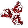

 PDBj
PDBj Assembly
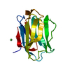


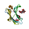
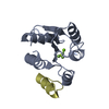
 Mass: 58.693 Da / Num. of mol.: 1 / Source method: obtained synthetically / Formula: Ni
 Mass: 18.015 Da / Num. of mol.: 180 / Source method: isolated from a natural source / Formula: H2O
 Sample preparation
 / Beamline: AR-NW12A / Wavelength: 1 Å
 Processing