 Movie
Movie Controller

[English] 日本語
 Yorodumi
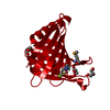
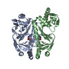
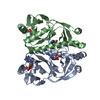
- PDB-3nv2: Crystal structure of human galectin-9 C-terminal CRD in complex w... -
+ Open data

- Basic information
| Entry | Database: PDB / ID: 3nv2 | |||||||||
|---|---|---|---|---|---|---|---|---|---|---|
| Title | Crystal structure of human galectin-9 C-terminal CRD in complex with N-acetyllactosamine | |||||||||
 Components Components | Galectin 9 short isoform variant | |||||||||
 Keywords Keywords | SUGAR BINDING PROTEIN / Sugar binding | |||||||||
| Function / homology |  Function and homology information Function and homology informationpositive regulation of activated T cell autonomous cell death / : / Interleukin-2 family signaling / positive regulation of dendritic cell apoptotic process / positive regulation of T cell activation via T cell receptor contact with antigen bound to MHC molecule on antigen presenting cell / negative regulation of mast cell degranulation / natural killer cell tolerance induction / positive regulation of dendritic cell differentiation / positive regulation of dendritic cell chemotaxis / galactoside binding ...positive regulation of activated T cell autonomous cell death / : / Interleukin-2 family signaling / positive regulation of dendritic cell apoptotic process / positive regulation of T cell activation via T cell receptor contact with antigen bound to MHC molecule on antigen presenting cell / negative regulation of mast cell degranulation / natural killer cell tolerance induction / positive regulation of dendritic cell differentiation / positive regulation of dendritic cell chemotaxis / galactoside binding / negative regulation of T-helper 1 type immune response / positive regulation of transforming growth factor beta production / galactose binding / oligosaccharide binding / NK T cell activation / positive regulation of CD4-positive, alpha-beta T cell proliferation / negative regulation of chemokine production / negative regulation of CD4-positive, alpha-beta T cell proliferation / disaccharide binding / positive regulation of interleukin-13 production / positive regulation of monocyte chemotactic protein-1 production / negative regulation of natural killer cell mediated cytotoxicity / positive regulation of interleukin-4 production / negative regulation of activated T cell proliferation / p38MAPK cascade / negative regulation of type II interferon production / positive regulation of interleukin-10 production / maternal process involved in female pregnancy / negative regulation of tumor necrosis factor production / : / ERK1 and ERK2 cascade / positive regulation of T cell migration / positive regulation of interleukin-12 production / response to interleukin-1 / positive regulation of interleukin-8 production / positive regulation of interleukin-1 beta production / female pregnancy / cellular response to virus / cellular response to type II interferon / positive regulation of non-canonical NF-kappaB signal transduction / positive regulation of interleukin-6 production / chemotaxis / positive regulation of type II interferon production / positive regulation of tumor necrosis factor production / carbohydrate binding / extracellular matrix / response to lipopolysaccharide / positive regulation of viral entry into host cell / positive regulation of ERK1 and ERK2 cascade / positive regulation of canonical NF-kappaB signal transduction / inflammatory response / receptor ligand activity / negative regulation of gene expression / positive regulation of gene expression / enzyme binding / : / nucleus / cytosol / cytoplasm Similarity search - Function | |||||||||
| Biological species |  Homo sapiens (human) Homo sapiens (human) | |||||||||
| Method |  X-RAY DIFFRACTION / SYNCHROTRON / MOLECULAR REPLACEMENT / Resolution: 2.34 Å X-RAY DIFFRACTION / SYNCHROTRON / MOLECULAR REPLACEMENT / Resolution: 2.34 Å | |||||||||
 Authors Authors | Yoshida, H. / Kamitori, S. | |||||||||
 Citation Citation | Journal: J.Biol.Chem. / Year: 2010 Title: X-ray structures of human galectin-9 C-terminal domain in complexes with a biantennary oligosaccharide and sialyllactose Authors: Yoshida, H. / Teraoka, M. / Nishi, N. / Nakakita, S. / Nakamura, T. / Hirashima, M. / Kamitori, S. | |||||||||
| History |
|
- Structure visualization
| Structure viewer | Molecule: MolmilJmol/JSmol |
|---|
- Downloads & links
-Download
| PDBx/mmCIF format | 3nv2.cif.gz | 47 KB | Display | PDBx/mmCIF format |
|---|---|---|---|---|
| PDB format | pdb3nv2.ent.gz | 31 KB | Display | PDB format |
| PDBx/mmJSON format | 3nv2.json.gz | Tree view | PDBx/mmJSON format | |
| Others |  Other downloads Other downloads |
-Validation report
| Arichive directory | https://data.pdbj.org/pub/pdb/validation_reports/nv/3nv2ftp://data.pdbj.org/pub/pdb/validation_reports/nv/3nv2 | HTTPS FTP |
|---|
-Related structure data
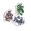
| Related structure data | 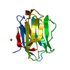 3nv1C 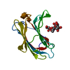 3nv3C 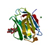 3nv4C  3koe C: citing same article ( S: Starting model for refinement |
|---|---|
| Similar structure data |


-Links
 PDBj
PDBj- Assembly
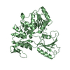
| Deposited unit | 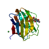
| ||||||||
|---|---|---|---|---|---|---|---|---|---|
| 1 | 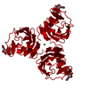
| ||||||||
| Unit cell |
| ||||||||
| Components on special symmetry positions |
| ||||||||
| Details | AUTHOR STATES THAT THE BIOLOGICAL ASSEMBLY IS UNKNOWN. |
-Components
| #1: Protein | Mass: 15729.982 Da / Num. of mol.: 1 Fragment: C-terminal carbohydrate recognition domain, residues 186-323 Source method: isolated from a genetically manipulated source Source: (gene. exp.) Homo sapiens (human) / Plasmid: pGEX-4T-2 / Production host:  |
|---|---|
| #2: Polysaccharide | beta-D-galactopyranose-(1-4)-2-acetamido-2-deoxy-alpha-D-glucopyranose / N-acetyl-alpha-lactosamine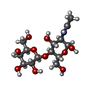  Source method: isolated from a genetically manipulated source Details: oligosaccharide / References: N-acetyl-alpha-lactosamine |
| #3: Chemical | ChemComp-NI /   Mass: 58.693 Da / Num. of mol.: 1 / Source method: obtained synthetically / Formula: Ni Mass: 58.693 Da / Num. of mol.: 1 / Source method: obtained synthetically / Formula: Ni |
| #4: Water | ChemComp-HOH /  Mass: 18.015 Da / Num. of mol.: 140 / Source method: isolated from a natural source / Formula: H2O Mass: 18.015 Da / Num. of mol.: 140 / Source method: isolated from a natural source / Formula: H2O |
| Nonpolymer details | THE LIGANDS GAL AND NDG FORM N-ACETYLLACT |
-Experimental details
-Experiment
| Experiment | Method: X-RAY DIFFRACTION / Number of used crystals: 1 |
|---|
- Sample preparation
| Crystal | Density Matthews: 2.31 Å3/Da / Density % sol: 46.75 % |
|---|---|
| Crystal grow | Temperature: 293 K / Method: vapor diffusion, hanging drop / pH: 8.5 Details: 20% PEG MME 2000, 0.1M Tris pH 8.5, 0.01M nickel chloride hexahydrate, VAPOR DIFFUSION, HANGING DROP, temperature 293K |
-Data collection
| Diffraction | Mean temperature: 100 K |
|---|---|
| Diffraction source | Source: SYNCHROTRON / Site: Photon Factory  / Beamline: AR-NW12A / Wavelength: 1 Å / Beamline: AR-NW12A / Wavelength: 1 Å |
| Detector | Type: ADSC QUANTUM 210r / Detector: CCD / Date: Nov 28, 2009 |
| Radiation | Protocol: SINGLE WAVELENGTH / Monochromatic (M) / Laue (L): M / Scattering type: x-ray |
| Radiation wavelength | Wavelength: 1 Å / Relative weight: 1 |
| Reflection | Resolution: 2.34→50 Å / Num. obs: 6135 / % possible obs: 100 % / Observed criterion σ(F): 0 / Observed criterion σ(I): 0 / Redundancy: 11.2 % / Biso Wilson estimate: 24.1 Å2 / Rmerge(I) obs: 0.075 / Net I/σ(I): 8.7 / Num. measured all: 68565 |
| Reflection shell | Resolution: 2.34→2.38 Å / Redundancy: 11.2 % / Rmerge(I) obs: 0.385 / Mean I/σ(I) obs: 7.4 / % possible all: 100 |
- Processing
| Software |
| |||||||||||||||||||||||||
|---|---|---|---|---|---|---|---|---|---|---|---|---|---|---|---|---|---|---|---|---|---|---|---|---|---|---|
| Refinement | Method to determine structure: MOLECULAR REPLACEMENT Starting model: PDB entry 3KOE 3koe Resolution: 2.34→38.85 Å / Rfactor Rfree error: 0.009 / Data cutoff high absF: 40669.38 / Data cutoff low absF: 0 / Isotropic thermal model: RESTRAINED / Cross valid method: THROUGHOUT / σ(F): 0 / Stereochemistry target values: Engh & Huber
| |||||||||||||||||||||||||
| Solvent computation | Solvent model: FLAT MODEL / Bsol: 60.142 Å2 / ksol: 0.364624 e/Å3 | |||||||||||||||||||||||||
| Displacement parameters |
| |||||||||||||||||||||||||
| Refine analyze |
| |||||||||||||||||||||||||
| Refinement step | Cycle: LAST / Resolution: 2.34→38.85 Å
| |||||||||||||||||||||||||
| Refine LS restraints |
| |||||||||||||||||||||||||
| LS refinement shell | Resolution: 2.34→2.49 Å / Rfactor Rfree error: 0.025 / Total num. of bins used: 6
| |||||||||||||||||||||||||
| Xplor file |
|
