 Movie
Movie Controller

[English] 日本語
 Yorodumi
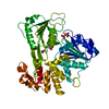
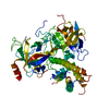
- PDB-2bh2: Crystal Structure of E. coli 5-methyluridine methyltransferase Ru... -
+ Open data

- Basic information
| Entry | Database: PDB / ID: 2bh2 | ||||||
|---|---|---|---|---|---|---|---|
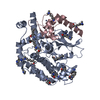
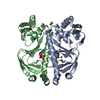
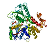
| Title | Crystal Structure of E. coli 5-methyluridine methyltransferase RumA in complex with ribosomal RNA substrate and S-adenosylhomocysteine. | ||||||
 Components Components |
| ||||||
 Keywords Keywords | TRANSFERASE / IRON-SULFUR CLUSTER / METHYLTRANSFERASE / RNA MODIFICATION / RNA PROCESSING / RUMA / BASE FLIPPING / SAM / OB-FOLD / PROTEIN-RNA COMPLEX / BASE STACKING / SUBSTRATE SELECTIVITY / GENERAL BASE / PRODUCT RELEASE / 4FE-4S / METAL-BINDING | ||||||
| Function / homology |  Function and homology information Function and homology information23S rRNA (uracil1939-C5)-methyltransferase / rRNA (uridine-C5-)-methyltransferase activity / rRNA base methylation / 4 iron, 4 sulfur cluster binding / iron ion binding / RNA binding Similarity search - Function | ||||||
| Biological species |  | ||||||
| Method |  X-RAY DIFFRACTION / SYNCHROTRON / MOLECULAR REPLACEMENT / Resolution: 2.15 Å X-RAY DIFFRACTION / SYNCHROTRON / MOLECULAR REPLACEMENT / Resolution: 2.15 Å | ||||||
 Authors Authors | Lee, T.T. / Agarwalla, S. / Stroud, R.M. | ||||||
 Citation Citation | Journal: Cell(Cambridge,Mass.) / Year: 2005 Title: A Unique RNA Fold in the Ruma-RNA-Cofactor Ternary Complex Contributes to Substrate Selectivity and Enzymatic Function Authors: Lee, T.T. / Agarwalla, S. / Stroud, R.M. #1: Journal: Structure / Year: 2004Title: Crystal Structure of Ruma, an Iron-Sulfur Cluster Containing E. Coli Ribosomal RNA 5-Methyluridine Methyltransferase Authors: Lee, T.T. / Agarwalla, S. / Stroud, R.M. | ||||||
| History |
|
- Structure visualization
| Structure viewer | Molecule: MolmilJmol/JSmol |
|---|
- Downloads & links
-Download
| PDBx/mmCIF format | 2bh2.cif.gz | 218.6 KB | Display | PDBx/mmCIF format |
|---|---|---|---|---|
| PDB format | pdb2bh2.ent.gz | 171.5 KB | Display | PDB format |
| PDBx/mmJSON format | 2bh2.json.gz | Tree view | PDBx/mmJSON format | |
| Others |  Other downloads Other downloads |
-Validation report
| Arichive directory | https://data.pdbj.org/pub/pdb/validation_reports/bh/2bh2ftp://data.pdbj.org/pub/pdb/validation_reports/bh/2bh2 | HTTPS FTP |
|---|
-Related structure data
| Related structure data | 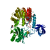 1uwvS S: Starting model for refinement |
|---|---|
| Similar structure data |

-Links
 PDBj
PDBj


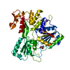
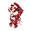
- Assembly
| Deposited unit | 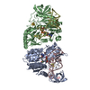
| ||||||||
|---|---|---|---|---|---|---|---|---|---|
| 1 | 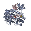
| ||||||||
| 2 | 
| ||||||||
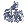
| Unit cell |
|
-Components
| #1: Protein | Mass: 48114.137 Da / Num. of mol.: 2 Source method: isolated from a genetically manipulated source Details: IRON-SULFUR CLUSTER LINKED BY CYS81, CYS87, CYS90, AND CYS162 Source: (gene. exp.) References: UniProt: P55135, Transferases; Transferring one-carbon groups; Methyltransferases #2: RNA chain | Mass: 11882.091 Da / Num. of mol.: 2 / Source method: obtained synthetically Details: 5-FLUORO-U1939 IS METHYLATED AND ITS 6-C IS COVALENTLY LINKED TO CYS389 OF RUMA Source: (synth.) #3: Chemical |   Mass: 384.411 Da / Num. of mol.: 2 / Source method: obtained synthetically / Formula: C14H20N6O5S Mass: 384.411 Da / Num. of mol.: 2 / Source method: obtained synthetically / Formula: C14H20N6O5S#4: Chemical |   Mass: 351.640 Da / Num. of mol.: 2 / Source method: obtained synthetically / Formula: Fe4S4 Mass: 351.640 Da / Num. of mol.: 2 / Source method: obtained synthetically / Formula: Fe4S4#5: Water | ChemComp-HOH / |  Mass: 18.015 Da / Num. of mol.: 340 / Source method: isolated from a natural source / Formula: H2O Mass: 18.015 Da / Num. of mol.: 340 / Source method: isolated from a natural source / Formula: H2OCompound details | S-ADENOSYL-L-METHIONINE => S-ADENOSYL-L-HOMOCYSTEINE + RRNA + RRNA CONTAINING THYMINE. CATALYZES ...S-ADENOSYL-L-METHIONINE | Has protein modification | Y | |
|---|
-Experimental details
-Experiment
| Experiment | Method: X-RAY DIFFRACTION / Number of used crystals: 1 |
|---|
- Sample preparation
| Crystal | Density Matthews: 2.3 Å3/Da / Density % sol: 47 % |
|---|---|
| Crystal grow | pH: 6.5 Details: RUMA-RNA-SAH COMPLEX WAS CRYSTALLIZED FROM 100 MM SODIUM CACODYLATE, PH 6.5, 1.5 M AMMONIUM SULFATE, AND 10 MM MGCL2 |
-Data collection
| Diffraction | Mean temperature: 100 K |
|---|---|
| Diffraction source | Source: SYNCHROTRON / Site: ALS  / Beamline: 8.3.1 / Wavelength: 1.1 / Beamline: 8.3.1 / Wavelength: 1.1 |
| Detector | Type: ADSC CCD / Detector: CCD / Date: Nov 19, 2003 |
| Radiation | Monochromator: DOUBLE CRYSTAL / Protocol: SINGLE WAVELENGTH / Monochromatic (M) / Laue (L): M / Scattering type: x-ray |
| Radiation wavelength | Wavelength: 1.1 Å / Relative weight: 1 |
| Reflection | Resolution: 2.15→50 Å / Num. obs: 59411 / % possible obs: 99.9 % / Observed criterion σ(I): 1.9 / Redundancy: 3.6 % / Rmerge(I) obs: 0.09 / Net I/σ(I): 12.7 |
| Reflection shell | Resolution: 2.15→2.19 Å / Redundancy: 3.7 % / Rmerge(I) obs: 0.73 / Mean I/σ(I) obs: 1.9 / % possible all: 100 |
- Processing
| Software |
| ||||||||||||||||||||||||||||||||||||||||||||||||||||||||||||||||||||||||||||||||||||||||||||||||||||||||||||||||||||||||||||||||||||||||||||||||||||||||||||||||||||||||||||||||||||||
|---|---|---|---|---|---|---|---|---|---|---|---|---|---|---|---|---|---|---|---|---|---|---|---|---|---|---|---|---|---|---|---|---|---|---|---|---|---|---|---|---|---|---|---|---|---|---|---|---|---|---|---|---|---|---|---|---|---|---|---|---|---|---|---|---|---|---|---|---|---|---|---|---|---|---|---|---|---|---|---|---|---|---|---|---|---|---|---|---|---|---|---|---|---|---|---|---|---|---|---|---|---|---|---|---|---|---|---|---|---|---|---|---|---|---|---|---|---|---|---|---|---|---|---|---|---|---|---|---|---|---|---|---|---|---|---|---|---|---|---|---|---|---|---|---|---|---|---|---|---|---|---|---|---|---|---|---|---|---|---|---|---|---|---|---|---|---|---|---|---|---|---|---|---|---|---|---|---|---|---|---|---|---|---|
| Refinement | Method to determine structure: MOLECULAR REPLACEMENT Starting model: PDB ENTRY 1UWV Resolution: 2.15→50 Å / Cor.coef. Fo:Fc: 0.96 / Cor.coef. Fo:Fc free: 0.932 / SU B: 5.547 / SU ML: 0.141 / Cross valid method: THROUGHOUT / ESU R: 0.261 / ESU R Free: 0.191 / Stereochemistry target values: MAXIMUM LIKELIHOOD / Details: HYDROGENS HAVE BEEN ADDED IN THE RIDING POSITIONS.
| ||||||||||||||||||||||||||||||||||||||||||||||||||||||||||||||||||||||||||||||||||||||||||||||||||||||||||||||||||||||||||||||||||||||||||||||||||||||||||||||||||||||||||||||||||||||
| Solvent computation | Ion probe radii: 0.8 Å / Shrinkage radii: 0.8 Å / VDW probe radii: 1.4 Å / Solvent model: BABINET MODEL WITH MASK | ||||||||||||||||||||||||||||||||||||||||||||||||||||||||||||||||||||||||||||||||||||||||||||||||||||||||||||||||||||||||||||||||||||||||||||||||||||||||||||||||||||||||||||||||||||||
| Displacement parameters | Biso mean: 27.19 Å2
| ||||||||||||||||||||||||||||||||||||||||||||||||||||||||||||||||||||||||||||||||||||||||||||||||||||||||||||||||||||||||||||||||||||||||||||||||||||||||||||||||||||||||||||||||||||||
| Refinement step | Cycle: LAST / Resolution: 2.15→50 Å
| ||||||||||||||||||||||||||||||||||||||||||||||||||||||||||||||||||||||||||||||||||||||||||||||||||||||||||||||||||||||||||||||||||||||||||||||||||||||||||||||||||||||||||||||||||||||
| Refine LS restraints |
|