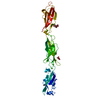
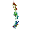
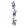
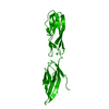
- PDB-3nr7: Crystal structure of S. typhimurium H-NS 1-83 -
+
Open data
ID or keywords:
Loading...
-
Basic information
Entry
Database: PDB / ID: 3nr7
Title
Crystal structure of S. typhimurium H-NS 1-83
Components
DNA-binding protein H-NS
Keywords
DNA BINDING PROTEIN / Dimer / oligomerisation / DNA condensation
Function / homology
Function and homology information
bent DNA binding / nucleoid / DNA-binding transcription repressor activity / negative regulation of gene expression, epigenetic / minor groove of adenine-thymine-rich DNA binding / protein-DNA complex / structural constituent of chromatin / protein dimerization activity / transcription cis-regulatory region binding / negative regulation of DNA-templated transcription ...bent DNA binding / nucleoid / DNA-binding transcription repressor activity / negative regulation of gene expression, epigenetic / minor groove of adenine-thymine-rich DNA binding / protein-DNA complex / structural constituent of chromatin / protein dimerization activity / transcription cis-regulatory region binding / negative regulation of DNA-templated transcription / DNA-templated transcription / DNA binding / identical protein binding / cytosol Similarity search - Function
H-NS histone-like proteins / Histone-like protein H-NS / Histone-like protein H-NS, N-terminal / : / H-NS histone-like proteins, N-terminal domain / Histone-like protein H-NS, C-terminal domain superfamily / Histone-like protein H-NS, C-terminal domain / H-NS histone C-terminal domain / Domain in histone-like proteins of HNS family / Helix Hairpins ...H-NS histone-like proteins / Histone-like protein H-NS / Histone-like protein H-NS, N-terminal / : / H-NS histone-like proteins, N-terminal domain / Histone-like protein H-NS, C-terminal domain superfamily / Histone-like protein H-NS, C-terminal domain / H-NS histone C-terminal domain / Domain in histone-like proteins of HNS family / Helix Hairpins / Orthogonal Bundle / Mainly Alpha Similarity search - Domain/homology
Resolution: 3.7→32.6 Å / Cor.coef. Fo:Fc: 0.939 / Cor.coef. Fo:Fc free: 0.93 / SU B: 119.068 / SU ML: 0.748 / Cross valid method: THROUGHOUT / ESU R Free: 0.578 / Stereochemistry target values: MAXIMUM LIKELIHOOD / Details: HYDROGENS HAVE BEEN ADDED IN THE RIDING POSITIONS
Rfactor
Num. reflection
% reflection
Selection details
Rfree
0.30375
427
7.5 %
RANDOM
Rwork
0.27107
-
-
-
all
0.27366
5676
-
-
obs
0.27366
5676
96.73 %
-
Solvent computation
Ion probe radii: 0.8 Å / Shrinkage radii: 0.8 Å / VDW probe radii: 1.4 Å / Solvent model: MASK
Displacement parameters
Biso mean: 189.256 Å2
Baniso -1
Baniso -2
Baniso -3
1-
5.23 Å2
2.62 Å2
-0 Å2
2-
-
5.23 Å2
-0 Å2
3-
-
-
-7.85 Å2
Refinement step
Cycle: LAST / Resolution: 3.7→32.6 Å
Protein
Nucleic acid
Ligand
Solvent
Total
Num. atoms
1288
0
0
0
1288
Refine LS restraints
Refine-ID
Type
Dev ideal
Dev ideal target
Number
X-RAY DIFFRACTION
r_bond_refined_d
0.014
0.022
1290
X-RAY DIFFRACTION
r_bond_other_d
X-RAY DIFFRACTION
r_angle_refined_deg
1.551
1.997
1732
X-RAY DIFFRACTION
r_angle_other_deg
X-RAY DIFFRACTION
r_dihedral_angle_1_deg
5.953
5
160
X-RAY DIFFRACTION
r_dihedral_angle_2_deg
44.592
25.143
70
X-RAY DIFFRACTION
r_dihedral_angle_3_deg
26.13
15
266
X-RAY DIFFRACTION
r_dihedral_angle_4_deg
21.975
15
16
X-RAY DIFFRACTION
r_chiral_restr
0.115
0.2
206
X-RAY DIFFRACTION
r_gen_planes_refined
0.004
0.02
952
X-RAY DIFFRACTION
r_gen_planes_other
X-RAY DIFFRACTION
r_nbd_refined
X-RAY DIFFRACTION
r_nbd_other
X-RAY DIFFRACTION
r_nbtor_refined
X-RAY DIFFRACTION
r_nbtor_other
X-RAY DIFFRACTION
r_xyhbond_nbd_refined
X-RAY DIFFRACTION
r_xyhbond_nbd_other
X-RAY DIFFRACTION
r_metal_ion_refined
X-RAY DIFFRACTION
r_metal_ion_other
X-RAY DIFFRACTION
r_symmetry_vdw_refined
X-RAY DIFFRACTION
r_symmetry_vdw_other
X-RAY DIFFRACTION
r_symmetry_hbond_refined
X-RAY DIFFRACTION
r_symmetry_hbond_other
X-RAY DIFFRACTION
r_symmetry_metal_ion_refined
X-RAY DIFFRACTION
r_symmetry_metal_ion_other
X-RAY DIFFRACTION
r_mcbond_it
0.377
1.5
808
X-RAY DIFFRACTION
r_mcbond_other
X-RAY DIFFRACTION
r_mcangle_it
0.733
2
1292
X-RAY DIFFRACTION
r_scbond_it
0.984
3
482
X-RAY DIFFRACTION
r_scangle_it
1.85
4.5
440
X-RAY DIFFRACTION
r_rigid_bond_restr
X-RAY DIFFRACTION
r_sphericity_free
X-RAY DIFFRACTION
r_sphericity_bonded
LS refinement shell
Resolution: 3.701→3.901 Å / Total num. of bins used: 10
Rfactor
Num. reflection
% reflection
Rfree
0.415
58
-
Rwork
0.413
740
-
obs
-
-
94.89 %
Refinement TLS params.
Method: refined / Refine-ID: X-RAY DIFFRACTION
ID
L11 (°2)
L12 (°2)
L13 (°2)
L22 (°2)
L23 (°2)
L33 (°2)
S11 (Å °)
S12 (Å °)
S13 (Å °)
S21 (Å °)
S22 (Å °)
S23 (Å °)
S31 (Å °)
S32 (Å °)
S33 (Å °)
T11 (Å2)
T12 (Å2)
T13 (Å2)
T22 (Å2)
T23 (Å2)
T33 (Å2)
Origin x (Å)
Origin y (Å)
Origin z (Å)
1
6.8763
-2.4733
10.6532
21.7359
-10.5618
16.9143
0.0247
1.6484
2.1055
2.6918
0.6817
-2.3914
-2.668
-0.2107
-0.7064
1.4069
0.0759
0.9436
1.0912
0.5563
2.2351
64.456
-22.123
6.507
2
20.1108
-4.4075
20.9768
17.0818
-6.9808
26.613
-0.8504
-0.5521
1.5358
-2.2025
0.2656
0.7736
-2.4228
-1.0372
0.5848
1.4109
0.0406
0.4313
0.5492
-0.0271
1.0768
64.614
-24.833
-7.95
3
17.7987
-4.2743
-12.1668
9.0261
-3.006
37.2198
1.0103
0.5069
-2.2873
-0.8312
0.7777
1.4837
0.999
-2.3182
-1.788
0.4088
-0.4976
0.1124
0.5763
-0.2457
1.1477
63.454
-34.214
-2.971
4
61.7193
1.6028
0.5027
18.8686
-1.8526
52.2527
1.2783
-2.3757
-1.358
2.1402
-0.431
-2.1995
-0.1697
2.654
-0.8473
0.3501
0.1583
-0.0813
0.3859
0.1656
0.7778
68.008
-32.112
5.005
5
91.1819
-0.0368
-8.0403
9.2158
0.9849
3.3776
3.3312
-1.398
-0.6907
-1.9346
-1.7786
-0.4707
1.8119
0.4551
-1.5526
1.5636
0.4344
0.2489
1.4338
-0.7774
1.4968
87.274
-41.265
-11.93
6
25.1985
6.0207
-7.1256
-0.8969
-4.3506
19.8422
1.2262
3.0186
0.4043
0.9533
0.3363
1.0016
-0.2606
-1.3938
-1.5626
0.8925
0.0083
1.0579
1.4868
0.2215
1.9079
44.992
-40.561
15.153
7
-2.3998
-16.7297
14.6929
32.2177
-7.1641
2.208
4.0199
-2.6903
-0.1467
-0.4507
-1.4838
-0.2426
-1.9465
0.4412
-2.5361
1.8807
-0.111
1.4379
1.8595
-0.5098
2.7685
104.885
-49.481
-14.723
8
0.3919
19.6174
-2.2413
19.2976
6.731
-3.8262
3.1538
3.3421
-1.3297
1.6461
-0.3399
-1.7311
1.3759
-0.2132
-2.814
1.4377
-0.2918
0.4466
2.9887
0.4586
2.0362
28.471
-50.677
18.969
9
-53.1194
-11.9535
-40.68
38.4964
4.1272
-18.6828
4.2818
0.236
-0.1528
-3.625
-0.5495
-3.7087
3.5776
1.1816
-3.7324
3.941
0.4667
-0.7981
3.5547
0.3276
3.4045
111.072
-43.759
-24.491
10
15.4838
-9.5522
5.6254
3.4537
-9.1023
0.6258
-1.1871
-1.5972
-1.3808
1.528
1.9588
0.1447
-1.5996
-1.808
-0.7716
1.7313
-0.1835
0.3532
2.1367
0.2216
2.8378
20.19
-43.804
25.489
Refinement TLS group
ID
Refine-ID
Refine TLS-ID
Auth asym-ID
Auth seq-ID
1
X-RAY DIFFRACTION
1
A
2 - 21
2
X-RAY DIFFRACTION
2
B
2 - 21
3
X-RAY DIFFRACTION
3
A
22 - 41
4
X-RAY DIFFRACTION
4
B
22 - 41
5
X-RAY DIFFRACTION
5
A
42 - 56
6
X-RAY DIFFRACTION
6
B
42 - 56
7
X-RAY DIFFRACTION
7
A
57 - 69
8
X-RAY DIFFRACTION
8
B
57 - 69
9
X-RAY DIFFRACTION
9
A
70 - 83
10
X-RAY DIFFRACTION
10
B
70 - 83
+
About Yorodumi
-
News
-
Feb 9, 2022. New format data for meta-information of EMDB entries
New format data for meta-information of EMDB entries
Version 3 of the EMDB header file is now the official format.
The previous official version 1.9 will be removed from the archive.
In the structure databanks used in Yorodumi, some data are registered as the other names, "COVID-19 virus" and "2019-nCoV". Here are the details of the virus and the list of structure data.
Jan 31, 2019. EMDB accession codes are about to change! (news from PDBe EMDB page)
EMDB accession codes are about to change! (news from PDBe EMDB page)
The allocation of 4 digits for EMDB accession codes will soon come to an end. Whilst these codes will remain in use, new EMDB accession codes will include an additional digit and will expand incrementally as the available range of codes is exhausted. The current 4-digit format prefixed with “EMD-” (i.e. EMD-XXXX) will advance to a 5-digit format (i.e. EMD-XXXXX), and so on. It is currently estimated that the 4-digit codes will be depleted around Spring 2019, at which point the 5-digit format will come into force.
The EM Navigator/Yorodumi systems omit the EMD- prefix.
Related info.:Q: What is EMD? / ID/Accession-code notation in Yorodumi/EM Navigator
Yorodumi is a browser for structure data from EMDB, PDB, SASBDB, etc.
This page is also the successor to EM Navigator detail page, and also detail information page/front-end page for Omokage search.
The word "yorodu" (or yorozu) is an old Japanese word meaning "ten thousand". "mi" (miru) is to see.
Related info.:EMDB / PDB / SASBDB / Comparison of 3 databanks / Yorodumi Search / Aug 31, 2016. New EM Navigator & Yorodumi / Yorodumi Papers / Jmol/JSmol / Function and homology information / Changes in new EM Navigator and Yorodumi
 Movie
Movie Controller


 Open data
 Basic information
 Components
 Keywords
 Function and homology information
 Salmonella enterica subsp. enterica serovar Typhimurium (bacteria)
 X-RAY DIFFRACTION /
  Authors
 Citation
 Structure visualization
 Downloads & links
 Other downloads


 PDBj
PDBj Assembly

 Sample preparation
 Processing