 Movie
Movie Controller

[English] 日本語
 Yorodumi
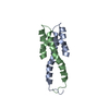
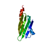
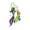
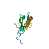
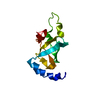
- PDB-1ov9: Crystal structure of the N-terminal dimerisation domain of VicH, ... -
+ Open data

- Basic information
| Entry | Database: PDB / ID: 1ov9 | ||||||
|---|---|---|---|---|---|---|---|
| Title | Crystal structure of the N-terminal dimerisation domain of VicH, the H-NS protein from Vibrio cholerae | ||||||
 Components Components | VicH protein | ||||||
 Keywords Keywords | DNA BINDING PROTEIN / dimer / helix / coiled-coil | ||||||
| Function / homology |  Function and homology information Function and homology informationbent DNA binding / nucleoid / DNA-binding transcription repressor activity / minor groove of adenine-thymine-rich DNA binding / protein-DNA complex / structural constituent of chromatin / protein dimerization activity / transcription cis-regulatory region binding / cytosol Similarity search - Function | ||||||
| Biological species |   Vibrio cholerae (bacteria) Vibrio cholerae (bacteria) | ||||||
| Method |  X-RAY DIFFRACTION / SYNCHROTRON / MOLECULAR REPLACEMENT / Resolution: 2.3 Å X-RAY DIFFRACTION / SYNCHROTRON / MOLECULAR REPLACEMENT / Resolution: 2.3 Å | ||||||
 Authors Authors | Cerdan, R. / Bloch, V. / Arold, S.T. | ||||||
 Citation Citation | Journal: J.Mol.Biol. / Year: 2003 Title: The Crystal Structure of the N-terminal dimerisation domain of VicH, the H-NS-like protein of Vibrio Cholerae Authors: Cerdan, R. / Bloch, V. / Yang, Y. / Bertin, P. / Dumas, C. / Rimsky, S. / Kochoyan, M. / Arold, S.T. | ||||||
| History |
|
- Structure visualization
| Structure viewer | Molecule: MolmilJmol/JSmol |
|---|
- Downloads & links
-Download
| PDBx/mmCIF format | 1ov9.cif.gz | 29.1 KB | Display | PDBx/mmCIF format |
|---|---|---|---|---|
| PDB format | pdb1ov9.ent.gz | 21 KB | Display | PDB format |
| PDBx/mmJSON format | 1ov9.json.gz | Tree view | PDBx/mmJSON format | |
| Others |  Other downloads Other downloads |
-Validation report
| Arichive directory | https://data.pdbj.org/pub/pdb/validation_reports/ov/1ov9ftp://data.pdbj.org/pub/pdb/validation_reports/ov/1ov9 | HTTPS FTP |
|---|
-Related structure data
| Related structure data | 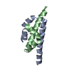 1ni8S S: Starting model for refinement |
|---|---|
| Similar structure data |


-Links
 PDBj
PDBj- Assembly
| Deposited unit | 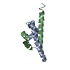
| |||||||||
|---|---|---|---|---|---|---|---|---|---|---|
| 1 |
| |||||||||
| Unit cell |
| |||||||||
| Components on special symmetry positions |
| |||||||||
| Details | the biological assembly is the dimer of the asymmetric unit. |
-Components
| #1: Protein/peptide | Mass: 5751.499 Da / Num. of mol.: 2 / Fragment: N-terminal Domain (residues 1-50) Source method: isolated from a genetically manipulated source Source: (gene. exp.) Vibrio cholerae (bacteria) / Gene: vich / Plasmid: pET22b / Species (production host): Escherichia coli / Production host: #2: Water | ChemComp-HOH / |  Mass: 18.015 Da / Num. of mol.: 47 / Source method: isolated from a natural source / Formula: H2O Mass: 18.015 Da / Num. of mol.: 47 / Source method: isolated from a natural source / Formula: H2O |
|---|
-Experimental details
-Experiment
| Experiment | Method: X-RAY DIFFRACTION / Number of used crystals: 1 |
|---|
- Sample preparation
| Crystal | Density Matthews: 2.07 Å3/Da / Density % sol: 40.02 % | ||||||||||||||||||||||||||||||||||||||||||
|---|---|---|---|---|---|---|---|---|---|---|---|---|---|---|---|---|---|---|---|---|---|---|---|---|---|---|---|---|---|---|---|---|---|---|---|---|---|---|---|---|---|---|---|
| Crystal grow | Temperature: 291 K / Method: vapor diffusion, hanging drop / pH: 7.5 Details: sodium chloride, PEG 3350, potassium iodide, pH 7.5, VAPOR DIFFUSION, HANGING DROP, temperature 291K | ||||||||||||||||||||||||||||||||||||||||||
| Crystal grow | *PLUS Temperature: 18 ℃ / Method: vapor diffusion, hanging drop | ||||||||||||||||||||||||||||||||||||||||||
| Components of the solutions | *PLUS
|
-Data collection
| Diffraction | Mean temperature: 100 K |
|---|---|
| Diffraction source | Source: SYNCHROTRON / Site: ESRF  / Beamline: BM14 / Wavelength: 0.9184 Å / Beamline: BM14 / Wavelength: 0.9184 Å |
| Detector | Type: ADSC QUANTUM 4 / Detector: CCD |
| Radiation | Monochromator: SAGITALLY FOCUSED Si(111) / Protocol: SINGLE WAVELENGTH / Monochromatic (M) / Laue (L): M / Scattering type: x-ray |
| Radiation wavelength | Wavelength: 0.9184 Å / Relative weight: 1 |
| Reflection | Resolution: 2.3→50.69 Å / Num. all: 3894 / Num. obs: 3894 / % possible obs: 88.8 % / Redundancy: 3.1 % / Biso Wilson estimate: 43 Å2 / Rmerge(I) obs: 0.1 / Rsym value: 0.085 / Net I/σ(I): 7 |
| Reflection shell | Resolution: 2.3→2.42 Å / Redundancy: 2.8 % / Rmerge(I) obs: 0.67 / Mean I/σ(I) obs: 1.2 / Num. unique all: 569 / Rsym value: 0.57 / % possible all: 90.8 |
| Reflection | *PLUS Lowest resolution: 50 Å / % possible obs: 90.8 % / Num. measured all: 11950 / Rmerge(I) obs: 0.085 |
| Reflection shell | *PLUS % possible obs: 90.8 % / Rmerge(I) obs: 0.57 |
- Processing
| Software |
| ||||||||||||||||||||||||||||
|---|---|---|---|---|---|---|---|---|---|---|---|---|---|---|---|---|---|---|---|---|---|---|---|---|---|---|---|---|---|
| Refinement | Method to determine structure: MOLECULAR REPLACEMENT Starting model: PDB entry 1NI8 Resolution: 2.3→50 Å / Isotropic thermal model: isotropic / Cross valid method: Maximum likelihood / σ(F): 0 / σ(I): 0 / Stereochemistry target values: Engh & Huber
| ||||||||||||||||||||||||||||
| Displacement parameters | Biso mean: 50 Å2 | ||||||||||||||||||||||||||||
| Refinement step | Cycle: LAST / Resolution: 2.3→50 Å
| ||||||||||||||||||||||||||||
| Refine LS restraints |
| ||||||||||||||||||||||||||||
| LS refinement shell | Resolution: 2.3→2.39 Å
| ||||||||||||||||||||||||||||
| Refine LS restraints | *PLUS Type: c_angle_d / Dev ideal: 1.4 |
