 Movie
Movie Controller


+ Open data

- Basic information
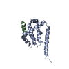
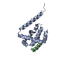
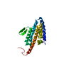
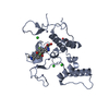
| Entry | Database: PDB / ID: 3npn | ||||||
|---|---|---|---|---|---|---|---|
| Title | Structure of the s-adenosylhomocysteine riboswitch at 3.0A | ||||||
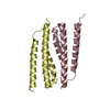
 Components Components | S-ADENOSYLHOMOCYSTEINE RIBOSWITCH | ||||||
 Keywords Keywords | RNA / riboswitch / S-adenosyl-homocysteine | ||||||
| Function / homology | S-ADENOSYL-L-HOMOCYSTEINE / RNA / RNA (> 10) Function and homology information Function and homology information | ||||||
| Biological species |  Ralstonia solanacearum (bacteria) Ralstonia solanacearum (bacteria) | ||||||
| Method |  X-RAY DIFFRACTION / SYNCHROTRON / SAD / Resolution: 2.792 Å X-RAY DIFFRACTION / SYNCHROTRON / SAD / Resolution: 2.792 Å | ||||||
 Authors Authors | Reyes, F.E. / Edwards, A.E. / Batey, R.T. | ||||||
 Citation Citation | Journal: Rna / Year: 2010 Title: Structural basis for recognition of S-adenosylhomocysteine by riboswitches. Authors: Edwards, A.L. / Reyes, F.E. / Heroux, A. / Batey, R.T. | ||||||
| History |
|
- Structure visualization
| Structure viewer | Molecule: MolmilJmol/JSmol |
|---|
- Downloads & links
-Download
| PDBx/mmCIF format | 3npn.cif.gz | 64.6 KB | Display | PDBx/mmCIF format |
|---|---|---|---|---|
| PDB format | pdb3npn.ent.gz | 49.9 KB | Display | PDB format |
| PDBx/mmJSON format | 3npn.json.gz | Tree view | PDBx/mmJSON format | |
| Others |  Other downloads Other downloads |
-Validation report
| Arichive directory | https://data.pdbj.org/pub/pdb/validation_reports/np/3npnftp://data.pdbj.org/pub/pdb/validation_reports/np/3npn | HTTPS FTP |
|---|
-Related structure data


-Links
 PDBj
PDBj


- Assembly
| Deposited unit | 
| ||||||||
|---|---|---|---|---|---|---|---|---|---|
| 1 |
| ||||||||
| Unit cell |
|
-Components
| #1: RNA chain | Mass: 17498.516 Da / Num. of mol.: 1 / Fragment: RNA / Source method: obtained synthetically Details: RNA was prepared by in vitro transcription with T7 RNA polymerase Source: (synth.) Ralstonia solanacearum (bacteria) |
|---|---|
| #2: Chemical | ChemComp-SAH /   Type: L-peptide linking / Mass: 384.411 Da / Num. of mol.: 1 / Source method: obtained synthetically / Formula: C14H20N6O5S Type: L-peptide linking / Mass: 384.411 Da / Num. of mol.: 1 / Source method: obtained synthetically / Formula: C14H20N6O5S |
-Experimental details
-Experiment
| Experiment | Method: X-RAY DIFFRACTION / Number of used crystals: 1 |
|---|
- Sample preparation
| Crystal | Density Matthews: 3.29 Å3/Da / Density % sol: 62.61 % |
|---|---|
| Crystal grow | Temperature: 298 K / Method: hanging drop / pH: 7 Details: 2.25 mM Spermine, 9 mM magnesium chloride, 0.9 mM spermidine, 1.8 mM Cobalt(III)hexammine chloride, 0.05 M sodium cacodylate pH 7.0, 5% (v/v) PEG 400, 20 mM iridium (III) hexammine chloride, ...Details: 2.25 mM Spermine, 9 mM magnesium chloride, 0.9 mM spermidine, 1.8 mM Cobalt(III)hexammine chloride, 0.05 M sodium cacodylate pH 7.0, 5% (v/v) PEG 400, 20 mM iridium (III) hexammine chloride, hanging drop, temperature 298K |
-Data collection
| Diffraction | Mean temperature: 100 K | |||||||||||||||||||||||||||||||||||||||||||||||||||||||||||||||||||||||||||||
|---|---|---|---|---|---|---|---|---|---|---|---|---|---|---|---|---|---|---|---|---|---|---|---|---|---|---|---|---|---|---|---|---|---|---|---|---|---|---|---|---|---|---|---|---|---|---|---|---|---|---|---|---|---|---|---|---|---|---|---|---|---|---|---|---|---|---|---|---|---|---|---|---|---|---|---|---|---|---|
| Diffraction source | Source: SYNCHROTRON / Site: NSLS  / Beamline: X25 / Wavelength: 1.1051 Å / Beamline: X25 / Wavelength: 1.1051 Å | |||||||||||||||||||||||||||||||||||||||||||||||||||||||||||||||||||||||||||||
| Detector | Type: ADSC QUANTUM 315 / Detector: CCD / Date: Jun 3, 2009 | |||||||||||||||||||||||||||||||||||||||||||||||||||||||||||||||||||||||||||||
| Radiation | Protocol: SINGLE WAVELENGTH / Monochromatic (M) / Laue (L): M / Scattering type: x-ray | |||||||||||||||||||||||||||||||||||||||||||||||||||||||||||||||||||||||||||||
| Radiation wavelength | Wavelength: 1.1051 Å / Relative weight: 1 | |||||||||||||||||||||||||||||||||||||||||||||||||||||||||||||||||||||||||||||
| Reflection | Resolution: 2.775→29.059 Å / Num. all: 5865 / Num. obs: 5854 / % possible obs: 99.8 % / Redundancy: 5.6 % / Rsym value: 0.123 / Net I/σ(I): 4.058 | |||||||||||||||||||||||||||||||||||||||||||||||||||||||||||||||||||||||||||||
| Reflection shell | Rmerge(I) obs: 0.01
|
-Phasing
| Phasing | Method: SAD | |||||||||||||||||||||||||||||||||||||||||||||||||
|---|---|---|---|---|---|---|---|---|---|---|---|---|---|---|---|---|---|---|---|---|---|---|---|---|---|---|---|---|---|---|---|---|---|---|---|---|---|---|---|---|---|---|---|---|---|---|---|---|---|---|
| Phasing MAD | D res high: 2.78 Å / D res low: 30 Å / FOM : 0.498 / FOM acentric: 0.518 / FOM centric: 0.332 / Reflection: 5849 / Reflection acentric: 5194 / Reflection centric: 651 | |||||||||||||||||||||||||||||||||||||||||||||||||
| Phasing dm | FOM : 0.67 / FOM acentric: 0.67 / FOM centric: 0.63 / Reflection: 5850 / Reflection acentric: 5199 / Reflection centric: 651 | |||||||||||||||||||||||||||||||||||||||||||||||||
| Phasing dm shell |
|
- Processing
| Software |
| |||||||||||||||||||||||||||||||||||||||||||||||||||||||||||||||||||||||||||||||||||||||||||||||||||||||||||||||||||||||||||||||||||||||||||||||||||
|---|---|---|---|---|---|---|---|---|---|---|---|---|---|---|---|---|---|---|---|---|---|---|---|---|---|---|---|---|---|---|---|---|---|---|---|---|---|---|---|---|---|---|---|---|---|---|---|---|---|---|---|---|---|---|---|---|---|---|---|---|---|---|---|---|---|---|---|---|---|---|---|---|---|---|---|---|---|---|---|---|---|---|---|---|---|---|---|---|---|---|---|---|---|---|---|---|---|---|---|---|---|---|---|---|---|---|---|---|---|---|---|---|---|---|---|---|---|---|---|---|---|---|---|---|---|---|---|---|---|---|---|---|---|---|---|---|---|---|---|---|---|---|---|---|---|---|---|---|
| Refinement | Method to determine structure: SAD / Resolution: 2.792→29.059 Å / Cor.coef. Fo:Fc: 0.93 / Cor.coef. Fo:Fc free: 0.9 / WRfactor Rfree: 0.302 / WRfactor Rwork: 0.265 / Occupancy max: 1 / Occupancy min: 1 / SU B: 36.426 / SU ML: 0.322 / Cross valid method: THROUGHOUT / σ(F): 0 / ESU R: 0.885 / ESU R Free: 0.392 / Stereochemistry target values: MAXIMUM LIKELIHOOD Details: HYDROGENS HAVE BEEN ADDED IN THE RIDING POSITIONS U VALUES : RESIDUAL ONLY
| |||||||||||||||||||||||||||||||||||||||||||||||||||||||||||||||||||||||||||||||||||||||||||||||||||||||||||||||||||||||||||||||||||||||||||||||||||
| Solvent computation | Ion probe radii: 0.8 Å / Shrinkage radii: 0.8 Å / VDW probe radii: 1.4 Å / Solvent model: MASK | |||||||||||||||||||||||||||||||||||||||||||||||||||||||||||||||||||||||||||||||||||||||||||||||||||||||||||||||||||||||||||||||||||||||||||||||||||
| Displacement parameters | Biso max: 88.54 Å2 / Biso mean: 78.29 Å2 / Biso min: 41.97 Å2
| |||||||||||||||||||||||||||||||||||||||||||||||||||||||||||||||||||||||||||||||||||||||||||||||||||||||||||||||||||||||||||||||||||||||||||||||||||
| Refinement step | Cycle: LAST / Resolution: 2.792→29.059 Å
| |||||||||||||||||||||||||||||||||||||||||||||||||||||||||||||||||||||||||||||||||||||||||||||||||||||||||||||||||||||||||||||||||||||||||||||||||||
| Refine LS restraints |
| |||||||||||||||||||||||||||||||||||||||||||||||||||||||||||||||||||||||||||||||||||||||||||||||||||||||||||||||||||||||||||||||||||||||||||||||||||
| LS refinement shell | Refine-ID: X-RAY DIFFRACTION / Total num. of bins used: 20
| |||||||||||||||||||||||||||||||||||||||||||||||||||||||||||||||||||||||||||||||||||||||||||||||||||||||||||||||||||||||||||||||||||||||||||||||||||
| Refinement TLS params. | Method: refined / Origin x: 122.7474 Å / Origin y: 68.5031 Å / Origin z: -4.2619 Å
|
