 Movie
Movie Controller

[English] 日本語
 Yorodumi
- PDB-3ng6: The crystal structure of hemoglobin I from Trematomus newnesi in ... -
+ Open data

- Basic information
| Entry | Database: PDB / ID: 3ng6 | ||||||
|---|---|---|---|---|---|---|---|
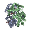
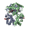
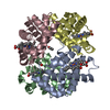
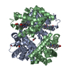
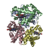
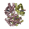
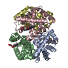
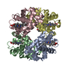
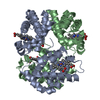
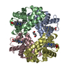
| Title | The crystal structure of hemoglobin I from Trematomus newnesi in deoxygenated state obtained through an oxidation/reduction cycle in which potassium hexacyanoferrate and sodium dithionite were alternatively added | ||||||
 Components Components |
| ||||||
 Keywords Keywords | OXYGEN TRANSPORT / Root effect / fish hemoglobin / antarctic fish | ||||||
| Function / homology |  Function and homology information Function and homology informationhemoglobin alpha binding / haptoglobin binding / haptoglobin-hemoglobin complex / organic acid binding / hemoglobin complex / hydrogen peroxide catabolic process / oxygen carrier activity / oxygen binding / peroxidase activity / blood microparticle ...hemoglobin alpha binding / haptoglobin binding / haptoglobin-hemoglobin complex / organic acid binding / hemoglobin complex / hydrogen peroxide catabolic process / oxygen carrier activity / oxygen binding / peroxidase activity / blood microparticle / iron ion binding / heme binding / metal ion binding Similarity search - Function | ||||||
| Biological species |  Trematomus newnesi (dusky notothen) Trematomus newnesi (dusky notothen) | ||||||
| Method |  X-RAY DIFFRACTION / MOLECULAR REPLACEMENT / Resolution: 2.2 Å X-RAY DIFFRACTION / MOLECULAR REPLACEMENT / Resolution: 2.2 Å | ||||||
 Authors Authors | Vergara, A. / Vitagliano, L. / Merlino, A. / Sica, F. / Marino, K. / Mazzarella, L. | ||||||
 Citation Citation | Journal: J.Biol.Chem. / Year: 2010 Title: An order-disorder transition plays a role in switching off the root effect in fish hemoglobins. Authors: Vergara, A. / Vitagliano, L. / Merlino, A. / Sica, F. / Marino, K. / Verde, C. / di Prisco, G. / Mazzarella, L. | ||||||
| History |
|
- Structure visualization
| Structure viewer | Molecule: MolmilJmol/JSmol |
|---|
- Downloads & links
-Download
| PDBx/mmCIF format | 3ng6.cif.gz | 129.4 KB | Display | PDBx/mmCIF format |
|---|---|---|---|---|
| PDB format | pdb3ng6.ent.gz | 101.9 KB | Display | PDB format |
| PDBx/mmJSON format | 3ng6.json.gz | Tree view | PDBx/mmJSON format | |
| Others |  Other downloads Other downloads |
-Validation report
| Summary document | 3ng6_validation.pdf.gz | 1.7 MB | Display | wwPDB validaton report |
|---|---|---|---|---|
| Full document | 3ng6_full_validation.pdf.gz | 1.7 MB | Display | |
| Data in XML | 3ng6_validation.xml.gz | 26.9 KB | Display | |
| Data in CIF | 3ng6_validation.cif.gz | 35 KB | Display | |
| Arichive directory | https://data.pdbj.org/pub/pdb/validation_reports/ng/3ng6ftp://data.pdbj.org/pub/pdb/validation_reports/ng/3ng6 | HTTPS FTP |
-Related structure data
| Related structure data |  3nfeC  2h8fS C: citing same article ( S: Starting model for refinement |
|---|---|
| Similar structure data |
-Links
 PDBj
PDBj


- Assembly
| Deposited unit | 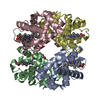
| ||||||||
|---|---|---|---|---|---|---|---|---|---|
| 1 |
| ||||||||
| Unit cell |
|
-Components
| #1: Protein | Mass: 15703.281 Da / Num. of mol.: 2 / Source method: isolated from a natural source / Source: (natural) Trematomus newnesi (dusky notothen) / References: UniProt: P45718#2: Protein | Mass: 16246.427 Da / Num. of mol.: 2 / Source method: isolated from a natural source / Source: (natural) Trematomus newnesi (dusky notothen) / References: UniProt: P45720#3: Chemical | ChemComp-HEM /   Mass: 616.487 Da / Num. of mol.: 4 / Source method: obtained synthetically / Formula: C34H32FeN4O4 Mass: 616.487 Da / Num. of mol.: 4 / Source method: obtained synthetically / Formula: C34H32FeN4O4#4: Water | ChemComp-HOH / |  Mass: 18.015 Da / Num. of mol.: 105 / Source method: isolated from a natural source / Formula: H2O Mass: 18.015 Da / Num. of mol.: 105 / Source method: isolated from a natural source / Formula: H2O |
|---|
-Experimental details
-Experiment
| Experiment | Method: X-RAY DIFFRACTION / Number of used crystals: 1 |
|---|
- Sample preparation
| Crystal | Density Matthews: 2.8 Å3/Da / Density % sol: 56.06 % |
|---|---|
| Crystal grow | Temperature: 293 K / Method: liquid diffusion / pH: 6 Details: protein at 10 mg/ml, in a 100 mM sodium acetate buffer pH 6.0, 2mM dithionite, poured into a capillary containing 20% (w/v) MPEG 5000 (2 mM dithionite), LIQUID DIFFUSION, temperature 293K |
-Data collection
| Diffraction | Mean temperature: 100 K |
|---|---|
| Diffraction source | Source: ROTATING ANODE / Type: RIGAKU MICROMAX-007 HF / Wavelength: 1.5418 Å |
| Detector | Type: ENRAF-NONIUS / Detector: CCD / Date: Jan 1, 2010 / Details: mirrors |
| Radiation | Monochromator: GRAPHITE / Protocol: SINGLE WAVELENGTH / Monochromatic (M) / Laue (L): M / Scattering type: x-ray |
| Radiation wavelength | Wavelength: 1.5418 Å / Relative weight: 1 |
| Reflection | Resolution: 2.2→26.6 Å / Num. obs: 32263 |
- Processing
| Software |
| ||||||||||||||||||||||
|---|---|---|---|---|---|---|---|---|---|---|---|---|---|---|---|---|---|---|---|---|---|---|---|
| Refinement | Method to determine structure: MOLECULAR REPLACEMENT Starting model: PDB entry 2H8F Resolution: 2.2→26.6 Å / Isotropic thermal model: Isotropic / Cross valid method: THROUGHOUT / Stereochemistry target values: Engh & Huber
| ||||||||||||||||||||||
| Refinement step | Cycle: LAST / Resolution: 2.2→26.6 Å
| ||||||||||||||||||||||
| Refine LS restraints |
|
