 Movie
Movie Controller

[English] 日本語
 Yorodumi
- PDB-3nfe: The crystal structure of hemoglobin I from trematomus newnesi in ... -
+ Open data

- Basic information
| Entry | Database: PDB / ID: 3nfe | ||||||
|---|---|---|---|---|---|---|---|
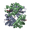
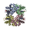
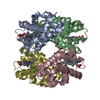
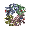
| Title | The crystal structure of hemoglobin I from trematomus newnesi in deoxygenated state | ||||||
 Components Components |
| ||||||
 Keywords Keywords | OXYGEN TRANSPORT / root effect / fish hemoglobin / antarctic fish | ||||||
| Function / homology |  Function and homology information Function and homology informationhaptoglobin binding / organic acid binding / haptoglobin-hemoglobin complex / hemoglobin complex / hydrogen peroxide catabolic process / oxygen carrier activity / peroxidase activity / oxygen binding / blood microparticle / iron ion binding ...haptoglobin binding / organic acid binding / haptoglobin-hemoglobin complex / hemoglobin complex / hydrogen peroxide catabolic process / oxygen carrier activity / peroxidase activity / oxygen binding / blood microparticle / iron ion binding / heme binding / metal ion binding Similarity search - Function | ||||||
| Biological species |  Trematomus newnesi (dusky notothen) Trematomus newnesi (dusky notothen) | ||||||
| Method |  X-RAY DIFFRACTION / MOLECULAR REPLACEMENT / Resolution: 2.01 Å X-RAY DIFFRACTION / MOLECULAR REPLACEMENT / Resolution: 2.01 Å | ||||||
 Authors Authors | Vergara, A. / Vitagliano, L. / Merlino, A. / Sica, F. / Marino, K. / Mazzarella, L. | ||||||
 Citation Citation | Journal: J.Biol.Chem. / Year: 2010 Title: An order-disorder transition plays a role in switching off the root effect in fish hemoglobins. Authors: Vergara, A. / Vitagliano, L. / Merlino, A. / Sica, F. / Marino, K. / Verde, C. / di Prisco, G. / Mazzarella, L. | ||||||
| History |
|
- Structure visualization
| Structure viewer | Molecule: MolmilJmol/JSmol |
|---|
- Downloads & links
-Download
| PDBx/mmCIF format | 3nfe.cif.gz | 129.1 KB | Display | PDBx/mmCIF format |
|---|---|---|---|---|
| PDB format | pdb3nfe.ent.gz | 102.2 KB | Display | PDB format |
| PDBx/mmJSON format | 3nfe.json.gz | Tree view | PDBx/mmJSON format | |
| Others |  Other downloads Other downloads |
-Validation report
| Arichive directory | https://data.pdbj.org/pub/pdb/validation_reports/nf/3nfeftp://data.pdbj.org/pub/pdb/validation_reports/nf/3nfe | HTTPS FTP |
|---|
-Related structure data
| Related structure data |  3ng6C  2h8fS C: citing same article ( S: Starting model for refinement |
|---|---|
| Similar structure data |


-Links
 PDBj
PDBj


- Assembly
| Deposited unit | 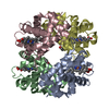
| ||||||||
|---|---|---|---|---|---|---|---|---|---|
| 1 |
| ||||||||
| Unit cell |
|
-Components
| #1: Protein | Mass: 15703.281 Da / Num. of mol.: 2 / Source method: isolated from a natural source / Source: (natural) Trematomus newnesi (dusky notothen) / References: UniProt: P45718#2: Protein | Mass: 16246.427 Da / Num. of mol.: 2 / Source method: isolated from a natural source / Source: (natural) Trematomus newnesi (dusky notothen) / References: UniProt: P45720#3: Chemical | ChemComp-HEM /   Mass: 616.487 Da / Num. of mol.: 4 / Source method: obtained synthetically / Formula: C34H32FeN4O4 Mass: 616.487 Da / Num. of mol.: 4 / Source method: obtained synthetically / Formula: C34H32FeN4O4#4: Water | ChemComp-HOH / |  Mass: 18.015 Da / Num. of mol.: 138 / Source method: isolated from a natural source / Formula: H2O Mass: 18.015 Da / Num. of mol.: 138 / Source method: isolated from a natural source / Formula: H2OHas protein modification | N | |
|---|
-Experimental details
-Experiment
| Experiment | Method: X-RAY DIFFRACTION / Number of used crystals: 1 |
|---|
- Sample preparation
| Crystal | Density Matthews: 2.83 Å3/Da / Density % sol: 56.47 % |
|---|---|
| Crystal grow | Temperature: 298 K / Method: liquid diffusion / pH: 6 Details: protein at 10 mg/ml, in 100 mM sodium acetate buffer pH 6.0, 2mM dithionite, poured into a capillary containing 20% (w/v) MPEG 5000 (2 mM dithionite), LIQUID DIFFUSION, temperature 298K |
-Data collection
| Diffraction | Mean temperature: 100 K |
|---|---|
| Diffraction source | Source: ROTATING ANODE / Type: RIGAKU MICROMAX-007 HF / Wavelength: 1.5418 Å |
| Detector | Type: ENRAF-NONIUS / Detector: CCD / Date: Jan 1, 2010 / Details: mirrors |
| Radiation | Monochromator: GRAPHITE / Protocol: SINGLE WAVELENGTH / Monochromatic (M) / Laue (L): M / Scattering type: x-ray |
| Radiation wavelength | Wavelength: 1.5418 Å / Relative weight: 1 |
| Reflection | Resolution: 2.01→39.7 Å / Num. obs: 42455 |
- Processing
| Software |
| ||||||||||||||||||||||
|---|---|---|---|---|---|---|---|---|---|---|---|---|---|---|---|---|---|---|---|---|---|---|---|
| Refinement | Method to determine structure: MOLECULAR REPLACEMENT Starting model: PDB entry 2H8F Resolution: 2.01→39.7 Å / Isotropic thermal model: Isotropic / Cross valid method: THROUGHOUT / σ(F): 0 / Stereochemistry target values: Engh & Huber Details: IN THE COURSE OF THE REFINEMENT (SEE BELOW), IT BECAME OBVIOUS THAT CRYSTALS WERE AFFECTED BY MEROHEDRAL TWINNING AND THE DIFFRACTION PATTERN WAS INTERPRETED AS RESULTING FROM TWO LATTICES ...Details: IN THE COURSE OF THE REFINEMENT (SEE BELOW), IT BECAME OBVIOUS THAT CRYSTALS WERE AFFECTED BY MEROHEDRAL TWINNING AND THE DIFFRACTION PATTERN WAS INTERPRETED AS RESULTING FROM TWO LATTICES CORRELATED BY ROTATION OF 180 AROUND AN AXIS PARALLEL TO A + B DIAGONAL. THEREFORE, THE INTENSITY ASSOCIATED TO EACH REFLECTION HKL IS THE WEIGHTED SUM OF TWO CONTRIBUTIONS: IO (HKL)= (1-C) I(HKL) + C I(KHL), C IS THE TWIN FRACTION, WHICH REFINED TO A VALUE OF 0.38
| ||||||||||||||||||||||
| Refinement step | Cycle: LAST / Resolution: 2.01→39.7 Å
| ||||||||||||||||||||||
| Refine LS restraints |
|
