 Movie
Movie Controller

[English] 日本語
 Yorodumi
- PDB-1la6: The crystal structure of Trematomus newnesi hemoglobin in a parti... -
+ Open data

- Basic information
| Entry | Database: PDB / ID: 1la6 | ||||||
|---|---|---|---|---|---|---|---|
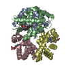
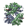
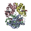
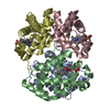
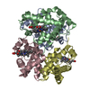
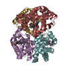
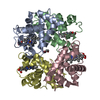
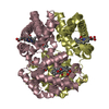
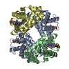
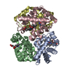
| Title | The crystal structure of Trematomus newnesi hemoglobin in a partial hemichrome state | ||||||
 Components Components |
| ||||||
 Keywords Keywords | OXYGEN STORAGE/TRANSPORT / Hemichrome / bishistidine complex / OXYGEN STORAGE-TRANSPORT COMPLEX | ||||||
| Function / homology |  Function and homology information Function and homology informationhaptoglobin binding / organic acid binding / haptoglobin-hemoglobin complex / hemoglobin complex / hydrogen peroxide catabolic process / oxygen carrier activity / peroxidase activity / oxygen binding / blood microparticle / iron ion binding ...haptoglobin binding / organic acid binding / haptoglobin-hemoglobin complex / hemoglobin complex / hydrogen peroxide catabolic process / oxygen carrier activity / peroxidase activity / oxygen binding / blood microparticle / iron ion binding / heme binding / metal ion binding Similarity search - Function | ||||||
| Biological species |  Trematomus newnesi (dusky notothen) Trematomus newnesi (dusky notothen) | ||||||
| Method |  X-RAY DIFFRACTION / MOLECULAR REPLACEMENT / Resolution: 2 Å X-RAY DIFFRACTION / MOLECULAR REPLACEMENT / Resolution: 2 Å | ||||||
 Authors Authors | Riccio, A. / Vitagliano, L. / di Prisco, G. / Zagari, A. / Mazzarella, L. | ||||||
 Citation Citation | Journal: Proc.Natl.Acad.Sci.USA / Year: 2002 Title: The crystal structure of a tetrameric hemoglobin in a partial hemichrome state Authors: Riccio, A. / Vitagliano, L. / di Prisco, G. / Zagari, A. / Mazzarella, L. #1: Journal: Acta Crystallogr. / Year: 2001Title: Liganded and unliganded forms of Antarctic fish haemoglobins in polyethylene glycol: crystallization of an R-state haemichrome intermediate Authors: Riccio, A. / Vitagliano, L. / di Prisco, G. / Zagari, A. / Mazzarella, L. #2: Journal: J.Mol.Biol. / Year: 1999Title: Crystal structure of Trematomus newnesi haemoglobin re-opens the root effect question Authors: Mazzarella, L. / D'Avino, R. / di PRISCO, G. / Savino, C. / VITAGLIANO, L. / MOODY, P.C.E. / ZAGARI, A. | ||||||
| History |
|
- Structure visualization
| Structure viewer | Molecule: MolmilJmol/JSmol |
|---|
- Downloads & links
-Download
| PDBx/mmCIF format | 1la6.cif.gz | 71.1 KB | Display | PDBx/mmCIF format |
|---|---|---|---|---|
| PDB format | pdb1la6.ent.gz | 51.9 KB | Display | PDB format |
| PDBx/mmJSON format | 1la6.json.gz | Tree view | PDBx/mmJSON format | |
| Others |  Other downloads Other downloads |
-Validation report
| Arichive directory | https://data.pdbj.org/pub/pdb/validation_reports/la/1la6ftp://data.pdbj.org/pub/pdb/validation_reports/la/1la6 | HTTPS FTP |
|---|
-Related structure data
| Related structure data | 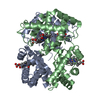 1t1nS S: Starting model for refinement |
|---|---|
| Similar structure data |
-Links
 PDBj
PDBj


- Assembly
| Deposited unit | 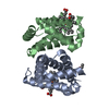
| ||||||||
|---|---|---|---|---|---|---|---|---|---|
| 1 | 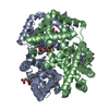
| ||||||||
| Unit cell |
| ||||||||
| Details | The biological assembly is a tetramer generated from the dimer in the asymmetric unit by the operation: -X, Y, -Z |
-Components
| #1: Protein | Mass: 15703.281 Da / Num. of mol.: 1 / Source method: isolated from a natural source / Source: (natural) Trematomus newnesi (dusky notothen) / References: UniProt: P45718 | ||||||
|---|---|---|---|---|---|---|---|
| #2: Protein | Mass: 16246.427 Da / Num. of mol.: 1 / Source method: isolated from a natural source / Source: (natural) Trematomus newnesi (dusky notothen) / References: UniProt: P45720 | ||||||
| #3: Chemical |   Mass: 616.487 Da / Num. of mol.: 2 / Source method: obtained synthetically / Formula: C34H32FeN4O4 Mass: 616.487 Da / Num. of mol.: 2 / Source method: obtained synthetically / Formula: C34H32FeN4O4#4: Chemical | ChemComp-CMO / |   Mass: 28.010 Da / Num. of mol.: 1 / Source method: obtained synthetically / Formula: CO Mass: 28.010 Da / Num. of mol.: 1 / Source method: obtained synthetically / Formula: CO#5: Water | ChemComp-HOH / |  Mass: 18.015 Da / Num. of mol.: 69 / Source method: isolated from a natural source / Formula: H2O Mass: 18.015 Da / Num. of mol.: 69 / Source method: isolated from a natural source / Formula: H2OHas protein modification | Y | |
-Experimental details
-Experiment
| Experiment | Method: X-RAY DIFFRACTION / Number of used crystals: 1 |
|---|
- Sample preparation
| Crystal | Density Matthews: 3.39 Å3/Da / Density % sol: 63.68 % | ||||||||||||||||||||||||
|---|---|---|---|---|---|---|---|---|---|---|---|---|---|---|---|---|---|---|---|---|---|---|---|---|---|
| Crystal grow | Temperature: 293 K / Method: free interface diffusion / pH: 7.6 Details: MPEG 5000, Sodium Acetate, pH 7.6, Free interface diffusion, temperature 293K | ||||||||||||||||||||||||
| Crystal grow | *PLUS pH: 6.2 / Details: Riccio, A., (2001) Acta Crystallogr., 57, 1144. | ||||||||||||||||||||||||
| Components of the solutions | *PLUS
|
-Data collection
| Diffraction | Mean temperature: 293 K |
|---|---|
| Diffraction source | Source: ROTATING ANODE / Type: ENRAF-NONIUS FR571 / Wavelength: 1.5418 Å |
| Detector | Type: MACSCIENCE / Detector: IMAGE PLATE / Date: Mar 30, 2000 / Details: mirrors |
| Radiation | Monochromator: mirrors / Protocol: SINGLE WAVELENGTH / Monochromatic (M) / Laue (L): M / Scattering type: x-ray |
| Radiation wavelength | Wavelength: 1.5418 Å / Relative weight: 1 |
| Reflection | Resolution: 2→12 Å / Num. all: 27020 / Num. obs: 27020 / % possible obs: 92.8 % / Observed criterion σ(I): -3 / Rmerge(I) obs: 0.077 |
| Reflection shell | Resolution: 2→2.15 Å / Rmerge(I) obs: 0.35 / % possible all: 87 |
| Reflection | *PLUS % possible obs: 92 % / Num. measured all: 64025 / Rmerge(I) obs: 0.077 |
| Reflection shell | *PLUS Rmerge(I) obs: 0.35 |
- Processing
| Software |
| ||||||||||||||||||||
|---|---|---|---|---|---|---|---|---|---|---|---|---|---|---|---|---|---|---|---|---|---|
| Refinement | Method to determine structure: MOLECULAR REPLACEMENT Starting model: PDB ENTRY 1T1N Resolution: 2→12 Å / Isotropic thermal model: Isotropic / Cross valid method: THROUGHOUT / σ(F): 2 / Stereochemistry target values: Engh & Huber
| ||||||||||||||||||||
| Refinement step | Cycle: LAST / Resolution: 2→12 Å
| ||||||||||||||||||||
| Refine LS restraints |
| ||||||||||||||||||||
| Refinement | *PLUS % reflection Rfree: 10 % / Rfactor all: 0.191 / Rfactor obs: 0.181 / Rfactor Rfree: 0.212 / Rfactor Rwork: 0.179 | ||||||||||||||||||||
| Solvent computation | *PLUS | ||||||||||||||||||||
| Displacement parameters | *PLUS | ||||||||||||||||||||
| Refine LS restraints | *PLUS
|
