 Movie
Movie Controller

[English] 日本語
 Yorodumi
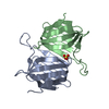
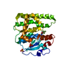
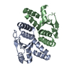
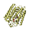
- PDB-3nct: X-ray crystal structure of the bacterial conjugation factor PsiB,... -
+ Open data

- Basic information
| Entry | Database: PDB / ID: 3nct | ||||||
|---|---|---|---|---|---|---|---|
| Title | X-ray crystal structure of the bacterial conjugation factor PsiB, a negative regulator of reca | ||||||
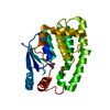
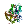
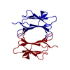
 Components Components | Protein psiB | ||||||
 Keywords Keywords | DNA BINDING PROTEIN / CHAPERONE | ||||||
| Function / homology | Plasmid SOS inhibition protein / Protein PsiB / Protein PsiB-like superfamily / Plasmid SOS inhibition protein (PsiB) / Rossmann fold / 3-Layer(aba) Sandwich / Alpha Beta / Protein PsiB Function and homology information Function and homology information | ||||||
| Biological species |  | ||||||
| Method |  X-RAY DIFFRACTION / SYNCHROTRON / SAD / Resolution: 2.2 Å X-RAY DIFFRACTION / SYNCHROTRON / SAD / Resolution: 2.2 Å | ||||||
 Authors Authors | Petrova, V. / Satyshur, K.A. / George, N.P. / McCaslin, D. / Cox, M.M. / Keck, J.L. | ||||||
 Citation Citation | Journal: J.Biol.Chem. / Year: 2010 Title: X-ray crystal structure of the bacterial conjugation factor PsiB, a negative regulator of RecA. Authors: Petrova, V. / Satyshur, K.A. / George, N.P. / McCaslin, D. / Cox, M.M. / Keck, J.L. | ||||||
| History |
|
- Structure visualization
| Structure viewer | Molecule: MolmilJmol/JSmol |
|---|
- Downloads & links
-Download
| PDBx/mmCIF format | 3nct.cif.gz | 229.6 KB | Display | PDBx/mmCIF format |
|---|---|---|---|---|
| PDB format | pdb3nct.ent.gz | 189.1 KB | Display | PDB format |
| PDBx/mmJSON format | 3nct.json.gz | Tree view | PDBx/mmJSON format | |
| Others |  Other downloads Other downloads |
-Validation report
| Arichive directory | https://data.pdbj.org/pub/pdb/validation_reports/nc/3nctftp://data.pdbj.org/pub/pdb/validation_reports/nc/3nct | HTTPS FTP |
|---|
-Related structure data
| Similar structure data |
|---|


-Links
 PDBj
PDBj
- Assembly
| Deposited unit | 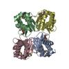
| ||||||||
|---|---|---|---|---|---|---|---|---|---|
| 1 | 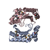
| ||||||||
| 2 | 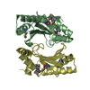
| ||||||||
| Unit cell |
|
-Components
| #1: Protein | Mass: 16127.248 Da / Num. of mol.: 4 Source method: isolated from a genetically manipulated source Source: (gene. exp.) #2: Water | ChemComp-HOH / |  Mass: 18.015 Da / Num. of mol.: 258 / Source method: isolated from a natural source / Formula: H2O Mass: 18.015 Da / Num. of mol.: 258 / Source method: isolated from a natural source / Formula: H2OHas protein modification | Y | Sequence details | THE AUTHORS CLAIM THAT THE UNIPROT ENTRY P10031 CONTAINS AN INCORRECT AMINOACID AT POSITION 137. ...THE AUTHORS CLAIM THAT THE UNIPROT ENTRY P10031 CONTAINS AN INCORRECT AMINOACID AT POSITION 137. THE CORRECT FULL SEQUENCE IS REPORTED IN THE PAPER BY DUTREIX M. ET AL. (NUCLEIC ACIDS RESEARCH 1988, V. 16, PAGES:10669-10679). SEE ALSO REMARK SEQADV. | |
|---|
-Experimental details
-Experiment
| Experiment | Method: X-RAY DIFFRACTION / Number of used crystals: 1 |
|---|
- Sample preparation
| Crystal | Density Matthews: 2.81 Å3/Da / Density % sol: 56.24 % |
|---|---|
| Crystal grow | Temperature: 298 K / Method: vapor diffusion, hanging drop / pH: 5.2 Details: 136 mM sodium acetate, pH 5.2, 1.4% PEG 4000, 20% glycerol, VAPOR DIFFUSION, HANGING DROP, temperature 298K |
-Data collection
| Diffraction | Mean temperature: 100 K |
|---|---|
| Diffraction source | Source: SYNCHROTRON / Site: APS  / Beamline: 21-ID-D / Wavelength: 0.97919 Å / Beamline: 21-ID-D / Wavelength: 0.97919 Å |
| Detector | Type: MARMOSAIC 300 mm CCD / Detector: CCD / Date: Apr 16, 2008 |
| Radiation | Monochromator: Si(111) / Protocol: SINGLE WAVELENGTH / Monochromatic (M) / Laue (L): M / Scattering type: x-ray |
| Radiation wavelength | Wavelength: 0.97919 Å / Relative weight: 1 |
| Reflection | Resolution: 2.2→30 Å / Num. obs: 71325 / % possible obs: 99.8 % / Observed criterion σ(F): 0 / Observed criterion σ(I): 0 / Redundancy: 6 % / Rmerge(I) obs: 0.059 / Net I/σ(I): 22 |
| Reflection shell | Resolution: 2.2→2.24 Å / Redundancy: 5.9 % / Rsym value: 0.269 / % possible all: 100 |
- Processing
| Software |
| |||||||||||||||||||||||||||||||||||||||||||||||||||||||||||||||||||||||||||||||||||||||||||||||||||||||||||||||||||||||||||||
|---|---|---|---|---|---|---|---|---|---|---|---|---|---|---|---|---|---|---|---|---|---|---|---|---|---|---|---|---|---|---|---|---|---|---|---|---|---|---|---|---|---|---|---|---|---|---|---|---|---|---|---|---|---|---|---|---|---|---|---|---|---|---|---|---|---|---|---|---|---|---|---|---|---|---|---|---|---|---|---|---|---|---|---|---|---|---|---|---|---|---|---|---|---|---|---|---|---|---|---|---|---|---|---|---|---|---|---|---|---|---|---|---|---|---|---|---|---|---|---|---|---|---|---|---|---|---|
| Refinement | Method to determine structure: SAD / Resolution: 2.2→30 Å / Cor.coef. Fo:Fc: 0.955 / Cor.coef. Fo:Fc free: 0.943 / SU B: 9.442 / SU ML: 0.111 / Cross valid method: THROUGHOUT / σ(F): 0 / ESU R Free: 0.169 / Stereochemistry target values: MAXIMUM LIKELIHOOD
| |||||||||||||||||||||||||||||||||||||||||||||||||||||||||||||||||||||||||||||||||||||||||||||||||||||||||||||||||||||||||||||
| Solvent computation | Ion probe radii: 0.8 Å / Shrinkage radii: 0.8 Å / VDW probe radii: 1.4 Å / Solvent model: BABINET MODEL WITH MASK | |||||||||||||||||||||||||||||||||||||||||||||||||||||||||||||||||||||||||||||||||||||||||||||||||||||||||||||||||||||||||||||
| Displacement parameters | Biso mean: 47.049 Å2
| |||||||||||||||||||||||||||||||||||||||||||||||||||||||||||||||||||||||||||||||||||||||||||||||||||||||||||||||||||||||||||||
| Refinement step | Cycle: LAST / Resolution: 2.2→30 Å
| |||||||||||||||||||||||||||||||||||||||||||||||||||||||||||||||||||||||||||||||||||||||||||||||||||||||||||||||||||||||||||||
| Refine LS restraints |
| |||||||||||||||||||||||||||||||||||||||||||||||||||||||||||||||||||||||||||||||||||||||||||||||||||||||||||||||||||||||||||||
| LS refinement shell | Resolution: 2.2→2.257 Å / Total num. of bins used: 20
| |||||||||||||||||||||||||||||||||||||||||||||||||||||||||||||||||||||||||||||||||||||||||||||||||||||||||||||||||||||||||||||
| Refinement TLS params. | Method: refined / Refine-ID: X-RAY DIFFRACTION
| |||||||||||||||||||||||||||||||||||||||||||||||||||||||||||||||||||||||||||||||||||||||||||||||||||||||||||||||||||||||||||||
| Refinement TLS group |
|
