 Movie
Movie Controller

[English] 日本語
 Yorodumi
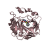
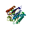
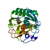
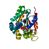
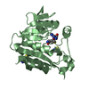
- PDB-3n7t: Crystal structure of a macrophage binding protein from Coccidioid... -
+ Open data

- Basic information
| Entry | Database: PDB / ID: 3n7t | ||||||
|---|---|---|---|---|---|---|---|
| Title | Crystal structure of a macrophage binding protein from Coccidioides immitis | ||||||
 Components Components | Macrophage binding protein | ||||||
 Keywords Keywords | PROTEIN BINDING / Seattle Structural Genomics Center for Infectious Disease / SSGCID / macrophage / pathogenic fungus / coccidioidomycosis / Valley Fever / meningitis | ||||||
| Function / homology |  Function and homology information Function and homology informationD-lactate dehydratase / glyoxalase III activity / methylglyoxal catabolic process to D-lactate via S-lactoyl-glutathione / cytoplasm Similarity search - Function | ||||||
| Biological species |  Coccidioides immitis (fungus) Coccidioides immitis (fungus) | ||||||
| Method |  X-RAY DIFFRACTION / SYNCHROTRON / MOLECULAR REPLACEMENT / molecular replacement / Resolution: 2.1 Å X-RAY DIFFRACTION / SYNCHROTRON / MOLECULAR REPLACEMENT / molecular replacement / Resolution: 2.1 Å | ||||||
 Authors Authors | Seattle Structural Genomics Center for Infectious Disease (SSGCID) | ||||||
 Citation Citation | Journal: TO BE PUBLISHED Title: Crystal structure of a macrophage binding protein from Coccidioides immitis Authors: Edwards, T.E. / Abendroth, J. / Sankaran, B. / Seattle Structural Genomics Center for Infectious Disease (SSGCID) | ||||||
| History |
|
- Structure visualization
| Structure viewer | Molecule: MolmilJmol/JSmol |
|---|
- Downloads & links
-Download
| PDBx/mmCIF format | 3n7t.cif.gz | 111.2 KB | Display | PDBx/mmCIF format |
|---|---|---|---|---|
| PDB format | pdb3n7t.ent.gz | 84.4 KB | Display | PDB format |
| PDBx/mmJSON format | 3n7t.json.gz | Tree view | PDBx/mmJSON format | |
| Others |  Other downloads Other downloads |
-Validation report
| Arichive directory | https://data.pdbj.org/pub/pdb/validation_reports/n7/3n7tftp://data.pdbj.org/pub/pdb/validation_reports/n7/3n7t | HTTPS FTP |
|---|
-Related structure data
| Related structure data |  1rw7 S: Starting model for refinement |
|---|---|
| Similar structure data | |
| Other databases |


-Links
 PDBj
PDBj

- Assembly
| Deposited unit | 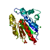
| ||||||||
|---|---|---|---|---|---|---|---|---|---|
| 1 |
| ||||||||
| 2 | 
| ||||||||
| Unit cell |
|
-Components
| #1: Protein | Mass: 27191.564 Da / Num. of mol.: 1 Source method: isolated from a genetically manipulated source Source: (gene. exp.) Coccidioides immitis (fungus) / Gene: MBP-1 / Plasmid: AVA0421 / Production host:  | ||||
|---|---|---|---|---|---|
| #2: Chemical | ChemComp-EDO /   Mass: 62.068 Da / Num. of mol.: 1 / Source method: obtained synthetically / Formula: C2H6O2 Mass: 62.068 Da / Num. of mol.: 1 / Source method: obtained synthetically / Formula: C2H6O2 | ||||
| #3: Chemical |   Mass: 35.453 Da / Num. of mol.: 3 / Source method: obtained synthetically / Formula: Cl Mass: 35.453 Da / Num. of mol.: 3 / Source method: obtained synthetically / Formula: Cl#4: Chemical | ChemComp-PO4 / |   Mass: 94.971 Da / Num. of mol.: 1 / Source method: obtained synthetically / Formula: PO4 Mass: 94.971 Da / Num. of mol.: 1 / Source method: obtained synthetically / Formula: PO4#5: Water | ChemComp-HOH / |  Mass: 18.015 Da / Num. of mol.: 232 / Source method: isolated from a natural source / Formula: H2O Mass: 18.015 Da / Num. of mol.: 232 / Source method: isolated from a natural source / Formula: H2O |
-Experimental details
-Experiment
| Experiment | Method: X-RAY DIFFRACTION / Number of used crystals: 1 |
|---|
- Sample preparation
| Crystal | Density Matthews: 3.93 Å3/Da / Density % sol: 68.72 % |
|---|---|
| Crystal grow | Temperature: 289 K / Method: vapor diffusion, sitting drop / pH: 4 Details: 32.1 mg/mL protein against JCSG+ screen condition C6, 40% PEG 300, 0.1 M phosphate/citrate pH 4.2, crystal tracking ID 206576c6, VAPOR DIFFUSION, SITTING DROP, temperature 289K |
-Data collection
| Diffraction | Mean temperature: 100 K |
|---|---|
| Diffraction source | Source: SYNCHROTRON / Site: ALS  / Beamline: 5.0.1 / Wavelength: 0.97946 Å / Beamline: 5.0.1 / Wavelength: 0.97946 Å |
| Detector | Type: ADSC QUANTUM 315r / Detector: CCD / Date: May 21, 2010 |
| Radiation | Protocol: SINGLE WAVELENGTH / Monochromatic (M) / Laue (L): M / Scattering type: x-ray |
| Radiation wavelength | Wavelength: 0.97946 Å / Relative weight: 1 |
| Reflection | Resolution: 2.1→50 Å / Num. obs: 25961 / % possible obs: 100 % / Observed criterion σ(I): -3 / Redundancy: 17.3 % / Biso Wilson estimate: 30.149 Å2 / Rmerge(I) obs: 0.104 / Net I/σ(I): 23.95 |
| Reflection shell | Resolution: 2.1→2.15 Å / Redundancy: 17.2 % / Rmerge(I) obs: 0.566 / Mean I/σ(I) obs: 6.1 / Num. measured obs: 32014 / Num. unique obs: 1863 / % possible all: 100 |
-Phasing
| Phasing | Method: molecular replacement | |||||||||
|---|---|---|---|---|---|---|---|---|---|---|
| Phasing MR | Rfactor: 52.23 / Model details: Phaser MODE: MR_AUTO
|
- Processing
| Software |
| |||||||||||||||||||||||||||||||||||||||||||||||||||||||||||||||||
|---|---|---|---|---|---|---|---|---|---|---|---|---|---|---|---|---|---|---|---|---|---|---|---|---|---|---|---|---|---|---|---|---|---|---|---|---|---|---|---|---|---|---|---|---|---|---|---|---|---|---|---|---|---|---|---|---|---|---|---|---|---|---|---|---|---|---|
| Refinement | Method to determine structure: MOLECULAR REPLACEMENT Starting model: PDB ENTRY 1rw7 1rw7 Resolution: 2.1→50 Å / Cor.coef. Fo:Fc: 0.96 / Cor.coef. Fo:Fc free: 0.936 / WRfactor Rfree: 0.18 / WRfactor Rwork: 0.148 / Occupancy max: 1 / Occupancy min: 0.5 / FOM work R set: 0.896 / SU B: 5.832 / SU ML: 0.071 / SU R Cruickshank DPI: 0.122 / SU Rfree: 0.12 / Cross valid method: THROUGHOUT / σ(F): 0 / ESU R Free: 0.12 / Stereochemistry target values: MAXIMUM LIKELIHOOD Details: HYDROGENS HAVE BEEN ADDED IN THE RIDING POSITIONS U VALUES : WITH TLS ADDED
| |||||||||||||||||||||||||||||||||||||||||||||||||||||||||||||||||
| Solvent computation | Ion probe radii: 0.8 Å / Shrinkage radii: 0.8 Å / VDW probe radii: 1.4 Å / Solvent model: MASK | |||||||||||||||||||||||||||||||||||||||||||||||||||||||||||||||||
| Displacement parameters | Biso max: 68.51 Å2 / Biso mean: 28.496 Å2 / Biso min: 14.49 Å2
| |||||||||||||||||||||||||||||||||||||||||||||||||||||||||||||||||
| Refinement step | Cycle: LAST / Resolution: 2.1→50 Å
| |||||||||||||||||||||||||||||||||||||||||||||||||||||||||||||||||
| Refine LS restraints |
| |||||||||||||||||||||||||||||||||||||||||||||||||||||||||||||||||
| LS refinement shell | Resolution: 2.1→2.154 Å / Total num. of bins used: 20
| |||||||||||||||||||||||||||||||||||||||||||||||||||||||||||||||||
| Refinement TLS params. | Method: refined / Origin x: 38.355 Å / Origin y: 16.0546 Å / Origin z: 41.1014 Å
|
