 Movie
Movie Controller


+ Open data

- Basic information
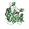
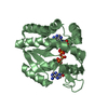
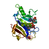
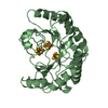
| Entry | Database: PDB / ID: 3bgd | ||||||
|---|---|---|---|---|---|---|---|
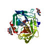
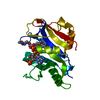
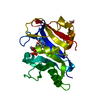
| Title | Thiopurine S-Methyltransferase | ||||||
 Components Components | Thiopurine S-methyltransferase | ||||||
 Keywords Keywords | TRANSFERASE / Methyltransferase / Cytoplasm / S-adenosyl-L-methionine | ||||||
| Function / homology |  Function and homology information Function and homology informationthiopurine S-methyltransferase / thiopurine S-methyltransferase activity / Methylation / Azathioprine ADME / S-adenosyl-L-methionine binding / xenobiotic metabolic process / methylation / cytoplasm Similarity search - Function | ||||||
| Biological species |  | ||||||
| Method |  X-RAY DIFFRACTION / SYNCHROTRON / MOLECULAR REPLACEMENT / molecular replacement / Resolution: 2 Å X-RAY DIFFRACTION / SYNCHROTRON / MOLECULAR REPLACEMENT / molecular replacement / Resolution: 2 Å | ||||||
 Authors Authors | Peng, Y. / Yee, V.C. | ||||||
 Citation Citation | Journal: Biochemistry / Year: 2008 Title: Structural Basis of Substrate Recognition in Thiopurine S-Methyltransferase Authors: Peng, Y. / Feng, Q. / Wilk, D. / Adjei, A.A. / Salavaggione, O.E. / Weinshilboum, R.M. / Yee, V.C. | ||||||
| History |
|
- Structure visualization
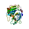
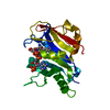
| Structure viewer | Molecule: MolmilJmol/JSmol |
|---|
- Downloads & links
-Download
| PDBx/mmCIF format | 3bgd.cif.gz | 114.3 KB | Display | PDBx/mmCIF format |
|---|---|---|---|---|
| PDB format | pdb3bgd.ent.gz | 88.2 KB | Display | PDB format |
| PDBx/mmJSON format | 3bgd.json.gz | Tree view | PDBx/mmJSON format | |
| Others |  Other downloads Other downloads |
-Validation report
| Arichive directory | https://data.pdbj.org/pub/pdb/validation_reports/bg/3bgdftp://data.pdbj.org/pub/pdb/validation_reports/bg/3bgd | HTTPS FTP |
|---|
-Related structure data
| Related structure data | 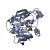 3bgiC  3bke 3bko C: citing same article ( |
|---|---|
| Similar structure data |
-Links
 PDBj
PDBj- Assembly
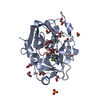
| Deposited unit | 
| ||||||||
|---|---|---|---|---|---|---|---|---|---|
| 1 | 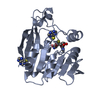
| ||||||||
| 2 | 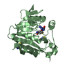
| ||||||||
| Unit cell |
|
-Components
| #1: Protein | Mass: 29796.074 Da / Num. of mol.: 2 Source method: isolated from a genetically manipulated source Source: (gene. exp.)  #2: Chemical |   Type: L-peptide linking / Mass: 384.411 Da / Num. of mol.: 2 / Source method: obtained synthetically / Formula: C14H20N6O5S Type: L-peptide linking / Mass: 384.411 Da / Num. of mol.: 2 / Source method: obtained synthetically / Formula: C14H20N6O5S#3: Chemical | ChemComp-PM6 / 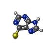  Mass: 152.177 Da / Num. of mol.: 4 / Source method: obtained synthetically / Formula: C5H4N4S / Comment: medication*YM Mass: 152.177 Da / Num. of mol.: 4 / Source method: obtained synthetically / Formula: C5H4N4S / Comment: medication*YM#4: Water | ChemComp-HOH / |  Mass: 18.015 Da / Num. of mol.: 307 / Source method: isolated from a natural source / Formula: H2O Mass: 18.015 Da / Num. of mol.: 307 / Source method: isolated from a natural source / Formula: H2O |
|---|
-Experimental details
-Experiment
| Experiment | Method: X-RAY DIFFRACTION / Number of used crystals: 1 |
|---|
- Sample preparation
| Crystal | Density Matthews: 2.39 Å3/Da / Density % sol: 48.64 % |
|---|---|
| Crystal grow | Temperature: 293 K / Method: vapor diffusion, sitting drop / pH: 5.5 Details: 20-22% PEG 3350, 0.15M malic acid, pH 5.5, VAPOR DIFFUSION, SITTING DROP, temperature 293.0K |
-Data collection
| Diffraction | Mean temperature: 100 K |
|---|---|
| Diffraction source | Source: SYNCHROTRON / Site: APS  / Beamline: 19-ID / Wavelength: 0.97912 Å / Beamline: 19-ID / Wavelength: 0.97912 Å |
| Detector | Type: ADSC QUANTUM 315 / Detector: CCD / Date: Mar 23, 2006 / Details: Mirrors |
| Radiation | Monochromator: Rosenbaum-Rock high-resolution double-crystal monochromator Protocol: SINGLE WAVELENGTH / Monochromatic (M) / Laue (L): M / Scattering type: x-ray |
| Radiation wavelength | Wavelength: 0.97912 Å / Relative weight: 1 |
| Reflection | Resolution: 2→30 Å / Num. all: 38056 / Num. obs: 36877 / % possible obs: 96.9 % / Observed criterion σ(F): 3 / Observed criterion σ(I): 3 / Redundancy: 2.9 % / Rsym value: 0.073 |
| Reflection shell | Resolution: 2→2.07 Å / Redundancy: 2.4 % / Mean I/σ(I) obs: 3.5 / Rsym value: 0.263 / % possible all: 83.6 |
-Phasing
| Phasing | Method: molecular replacement |
|---|
- Processing
| Software |
| ||||||||||||||||||||||||||||||||||||||||||||||||||||||||||||||||||||||||||||||||||||||||||||||||||||||||||||||||||||||||||||||||||||||||||||||||||||||||||||||||||||||||||
|---|---|---|---|---|---|---|---|---|---|---|---|---|---|---|---|---|---|---|---|---|---|---|---|---|---|---|---|---|---|---|---|---|---|---|---|---|---|---|---|---|---|---|---|---|---|---|---|---|---|---|---|---|---|---|---|---|---|---|---|---|---|---|---|---|---|---|---|---|---|---|---|---|---|---|---|---|---|---|---|---|---|---|---|---|---|---|---|---|---|---|---|---|---|---|---|---|---|---|---|---|---|---|---|---|---|---|---|---|---|---|---|---|---|---|---|---|---|---|---|---|---|---|---|---|---|---|---|---|---|---|---|---|---|---|---|---|---|---|---|---|---|---|---|---|---|---|---|---|---|---|---|---|---|---|---|---|---|---|---|---|---|---|---|---|---|---|---|---|---|---|---|
| Refinement | Method to determine structure: MOLECULAR REPLACEMENT / Resolution: 2→30 Å / Cor.coef. Fo:Fc: 0.928 / Cor.coef. Fo:Fc free: 0.892 / SU B: 4.796 / SU ML: 0.135 / Cross valid method: THROUGHOUT / ESU R: 0.204 / ESU R Free: 0.186 / Stereochemistry target values: MAXIMUM LIKELIHOOD / Details: HYDROGENS HAVE BEEN ADDED IN THE RIDING POSITIONS
| ||||||||||||||||||||||||||||||||||||||||||||||||||||||||||||||||||||||||||||||||||||||||||||||||||||||||||||||||||||||||||||||||||||||||||||||||||||||||||||||||||||||||||
| Solvent computation | Ion probe radii: 0.8 Å / Shrinkage radii: 0.8 Å / VDW probe radii: 1.2 Å / Solvent model: MASK | ||||||||||||||||||||||||||||||||||||||||||||||||||||||||||||||||||||||||||||||||||||||||||||||||||||||||||||||||||||||||||||||||||||||||||||||||||||||||||||||||||||||||||
| Displacement parameters | Biso mean: 24.613 Å2
| ||||||||||||||||||||||||||||||||||||||||||||||||||||||||||||||||||||||||||||||||||||||||||||||||||||||||||||||||||||||||||||||||||||||||||||||||||||||||||||||||||||||||||
| Refinement step | Cycle: LAST / Resolution: 2→30 Å
| ||||||||||||||||||||||||||||||||||||||||||||||||||||||||||||||||||||||||||||||||||||||||||||||||||||||||||||||||||||||||||||||||||||||||||||||||||||||||||||||||||||||||||
| Refine LS restraints |
| ||||||||||||||||||||||||||||||||||||||||||||||||||||||||||||||||||||||||||||||||||||||||||||||||||||||||||||||||||||||||||||||||||||||||||||||||||||||||||||||||||||||||||
| LS refinement shell | Resolution: 2→2.052 Å / Total num. of bins used: 20
|
