 Movie
Movie Controller

[English] 日本語
 Yorodumi
- PDB-2gb4: Crystal structure of Thiopurine methyltransferase (18204406) from... -
+ Open data

- Basic information
| Entry | Database: PDB / ID: 2gb4 | ||||||
|---|---|---|---|---|---|---|---|
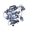
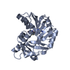
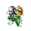
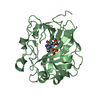
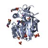
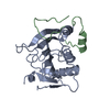
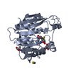
| Title | Crystal structure of Thiopurine methyltransferase (18204406) from Mus musculus at 1.35 A resolution | ||||||
 Components Components | Thiopurine S-methyltransferase | ||||||
 Keywords Keywords | TRANSFERASE / 18204406 / Thiopurine methyltransferase / Structural Genomics / PSI / Protein Structure Initiative / Joint Center for Structural Genomics / JCSG | ||||||
| Function / homology |  Function and homology information Function and homology informationthiopurine S-methyltransferase / thiopurine S-methyltransferase activity / Methylation / Azathioprine ADME / S-adenosyl-L-methionine binding / xenobiotic metabolic process / methylation / cytoplasm Similarity search - Function | ||||||
| Biological species |  | ||||||
| Method |  X-RAY DIFFRACTION / SYNCHROTRON / MOLECULAR REPLACEMENT / molecular replacement / Resolution: 1.25 Å X-RAY DIFFRACTION / SYNCHROTRON / MOLECULAR REPLACEMENT / molecular replacement / Resolution: 1.25 Å | ||||||
 Authors Authors | Joint Center for Structural Genomics (JCSG) | ||||||
 Citation Citation | Journal: To be published Title: Crystal structure of Thiopurine methyltransferase (18204406) from Mus musculus at 1.35 A resolution Authors: Joint Center for Structural Genomics (JCSG) | ||||||
| History |
|
- Structure visualization
| Structure viewer | Molecule: MolmilJmol/JSmol |
|---|
- Downloads & links
-Download
| PDBx/mmCIF format | 2gb4.cif.gz | 223.2 KB | Display | PDBx/mmCIF format |
|---|---|---|---|---|
| PDB format | pdb2gb4.ent.gz | 177.7 KB | Display | PDB format |
| PDBx/mmJSON format | 2gb4.json.gz | Tree view | PDBx/mmJSON format | |
| Others |  Other downloads Other downloads |
-Validation report
| Arichive directory | https://data.pdbj.org/pub/pdb/validation_reports/gb/2gb4ftp://data.pdbj.org/pub/pdb/validation_reports/gb/2gb4 | HTTPS FTP |
|---|
-Related structure data
| Related structure data | 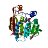 2bzgS S: Starting model for refinement |
|---|---|
| Similar structure data | |
| Other databases |


-Links
 PDBj
PDBj- Assembly
| Deposited unit | 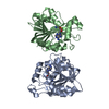
| ||||||||
|---|---|---|---|---|---|---|---|---|---|
| 1 | 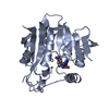
| ||||||||
| 2 | 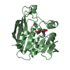
| ||||||||
| Unit cell |
|
-Components
| #1: Protein | Mass: 29086.346 Da / Num. of mol.: 2 Source method: isolated from a genetically manipulated source Source: (gene. exp.)  #2: Chemical |   Type: L-peptide linking / Mass: 384.411 Da / Num. of mol.: 2 / Source method: obtained synthetically / Formula: C14H20N6O5S Type: L-peptide linking / Mass: 384.411 Da / Num. of mol.: 2 / Source method: obtained synthetically / Formula: C14H20N6O5S#3: Water | ChemComp-HOH / |  Mass: 18.015 Da / Num. of mol.: 402 / Source method: isolated from a natural source / Formula: H2O Mass: 18.015 Da / Num. of mol.: 402 / Source method: isolated from a natural source / Formula: H2O |
|---|
-Experimental details
-Experiment
| Experiment | Method: X-RAY DIFFRACTION / Number of used crystals: 1 |
|---|
- Sample preparation
| Crystal | Density Matthews: 2.43 Å3/Da / Density % sol: 48.98 % |
|---|---|
| Crystal grow | Temperature: 293 K / Method: vapor diffusion, sitting drop, nanodrop / pH: 5.6 Details: 5.0% Glycerol, 19.0% iso-Propanol, 19.0% PEG-4000, 0.1M Citrate, pH 5.6, VAPOR DIFFUSION, SITTING DROP, NANODROP, temperature 293K |
-Data collection
| Diffraction | Mean temperature: 100 K | ||||||||||||||||||||||||||||||||||||||||||||
|---|---|---|---|---|---|---|---|---|---|---|---|---|---|---|---|---|---|---|---|---|---|---|---|---|---|---|---|---|---|---|---|---|---|---|---|---|---|---|---|---|---|---|---|---|---|
| Diffraction source | Source: SYNCHROTRON / Site: SSRL  / Beamline: BL9-2 / Wavelength: 1 / Beamline: BL9-2 / Wavelength: 1 | ||||||||||||||||||||||||||||||||||||||||||||
| Detector | Type: MARMOSAIC 325 mm CCD / Detector: CCD / Date: Jul 23, 2005 | ||||||||||||||||||||||||||||||||||||||||||||
| Radiation | Monochromator: Double crystal monochromator / Protocol: SINGLE WAVELENGTH / Monochromatic (M) / Laue (L): M / Scattering type: x-ray | ||||||||||||||||||||||||||||||||||||||||||||
| Radiation wavelength | Wavelength: 1 Å / Relative weight: 1 | ||||||||||||||||||||||||||||||||||||||||||||
| Reflection | Resolution: 1.25→50 Å / Num. obs: 125498 / % possible obs: 79.4 % / Redundancy: 2.7 % / Rmerge(I) obs: 0.033 / Net I/σ(I): 15.4 | ||||||||||||||||||||||||||||||||||||||||||||
| Reflection shell |
|
-Phasing
| Phasing | Method: molecular replacement |
|---|
- Processing
| Software |
| |||||||||||||||||||||||||||||||||
|---|---|---|---|---|---|---|---|---|---|---|---|---|---|---|---|---|---|---|---|---|---|---|---|---|---|---|---|---|---|---|---|---|---|---|
| Refinement | Method to determine structure: MOLECULAR REPLACEMENT Starting model: pdb entry 2bzg Resolution: 1.25→35.9 Å / Cor.coef. Fo:Fc: 0.935 / Cor.coef. Fo:Fc free: 0.925 / Num. parameters: 38290 / Num. restraintsaints: 50906 / SU B: 2.556 / SU ML: 0.059 / Cross valid method: FREE R / σ(F): 0 / ESU R: 0.07 / ESU R Free: 0.072 / Stereochemistry target values: MAXIMUM LIKELIHOOD Details: 1. HYDROGENS HAVE BEEN ADDED IN THE RIDING POSITIONS. 2. ANISOTROPIC REFINEMENT REDUCED FREE R (NO CUTOFF) BY 8%. 3. THE NOMINAL RESOLUTION IS 1.35A WITH 16569 REFLECTIONS BETWEEN 1.35-1.25A ...Details: 1. HYDROGENS HAVE BEEN ADDED IN THE RIDING POSITIONS. 2. ANISOTROPIC REFINEMENT REDUCED FREE R (NO CUTOFF) BY 8%. 3. THE NOMINAL RESOLUTION IS 1.35A WITH 16569 REFLECTIONS BETWEEN 1.35-1.25A (50.6% COMPLETE FOR THIS SHELL) INCLUDED IN THIS REFINEMENT. 4. REFINEMENT WAS AGAINST INTENSITY DATA. 5. NCS RESTRAINTS WERE APPLIED BETWEEN RESIDUES 40-158, 162-186 AND 205-240 OF CHAINS A AND B. 6. RESIDUES 1-9 OF CHAIN A AND 1-10 OF CHAIN B HAVE NO DENSITY AND WERE NOT MODELED. 7. S-ADENOSYL-L-HOMOCYSTEINE MODELED BASED ON ELECTRON DENSITY AND PROPOSED FUNCTION. 8. DATA WAS REFINED WITH TWIN LAW -H, -K, H+L, TWIN FRACTION 0.50. 9. THE RFREE SET WAS GENERATED WITH THE TWIN LAW.
| |||||||||||||||||||||||||||||||||
| Solvent computation | Solvent model: MOEWS & KRETSINGER, J.MOL.BIOL.91(1973)201-228 | |||||||||||||||||||||||||||||||||
| Displacement parameters | Biso mean: 18.394 Å2
| |||||||||||||||||||||||||||||||||
| Refine analyze | Num. disordered residues: 11 / Occupancy sum hydrogen: 3551 / Occupancy sum non hydrogen: 4169 | |||||||||||||||||||||||||||||||||
| Refinement step | Cycle: LAST / Resolution: 1.25→35.9 Å
| |||||||||||||||||||||||||||||||||
| Refine LS restraints |
| |||||||||||||||||||||||||||||||||
| LS refinement shell | Resolution: 1.249→1.282 Å / Total num. of bins used: 20
|
