 Movie
Movie Controller

[English] 日本語
 Yorodumi
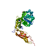
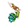
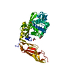
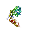
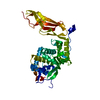
- PDB-3mzf: Structure of penicillin-binding protein 5 from E. coli: imipenem ... -
+ Open data

- Basic information
| Entry | Database: PDB / ID: 3mzf | ||||||
|---|---|---|---|---|---|---|---|
| Title | Structure of penicillin-binding protein 5 from E. coli: imipenem acyl-enzyme complex | ||||||
 Components Components | D-alanyl-D-alanine carboxypeptidase dacA | ||||||
 Keywords Keywords | HYDROLASE/ANTIBIOTIC / BETA-LACTAM ANTIBIOTIC / PENICILLIN-BINDING PROTEIN / DD-CARBOXYPEPTIDASE / HYDROLASE / HYDROLASE-ANTIBIOTIC complex | ||||||
| Function / homology |  Function and homology information Function and homology informationpeptidoglycan metabolic process / serine-type D-Ala-D-Ala carboxypeptidase / serine-type D-Ala-D-Ala carboxypeptidase activity / penicillin binding / peptidoglycan biosynthetic process / carboxypeptidase activity / cell wall organization / beta-lactamase activity / beta-lactamase / regulation of cell shape ...peptidoglycan metabolic process / serine-type D-Ala-D-Ala carboxypeptidase / serine-type D-Ala-D-Ala carboxypeptidase activity / penicillin binding / peptidoglycan biosynthetic process / carboxypeptidase activity / cell wall organization / beta-lactamase activity / beta-lactamase / regulation of cell shape / outer membrane-bounded periplasmic space / cell division / protein homodimerization activity / proteolysis / plasma membrane Similarity search - Function | ||||||
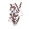
| Biological species |  | ||||||
| Method |  X-RAY DIFFRACTION / SYNCHROTRON / FOURIER SYNTHESIS / Resolution: 1.5 Å X-RAY DIFFRACTION / SYNCHROTRON / FOURIER SYNTHESIS / Resolution: 1.5 Å | ||||||
 Authors Authors | Nicola, G. / Tomberg, J. / Pratt, R.F. / Nicholas, R.A. / Davies, C. | ||||||
 Citation Citation | Journal: Biochemistry / Year: 2010 Title: Crystal structures of covalent complexes of beta-lactam antibiotics with Escherichia coli penicillin-binding protein 5: toward an understanding of antibiotic specificity Authors: Nicola, G. / Tomberg, J. / Pratt, R.F. / Nicholas, R.A. / Davies, C. | ||||||
| History |
|
- Structure visualization
| Structure viewer | Molecule: MolmilJmol/JSmol |
|---|
- Downloads & links
-Download
| PDBx/mmCIF format | 3mzf.cif.gz | 167.2 KB | Display | PDBx/mmCIF format |
|---|---|---|---|---|
| PDB format | pdb3mzf.ent.gz | 131.7 KB | Display | PDB format |
| PDBx/mmJSON format | 3mzf.json.gz | Tree view | PDBx/mmJSON format | |
| Others |  Other downloads Other downloads |
-Validation report
| Arichive directory | https://data.pdbj.org/pub/pdb/validation_reports/mz/3mzfftp://data.pdbj.org/pub/pdb/validation_reports/mz/3mzf | HTTPS FTP |
|---|
-Related structure data
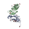
| Related structure data |  3mzdC 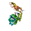 3mzeC 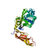 1nzoS S: Starting model for refinement C: citing same article ( |
|---|---|
| Similar structure data |


-Links
 PDBj
PDBj
- Assembly
| Deposited unit | 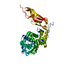
| ||||||||
|---|---|---|---|---|---|---|---|---|---|
| 1 |
| ||||||||
| Unit cell |
|
-Components
| #1: Protein | Mass: 39841.117 Da / Num. of mol.: 1 / Fragment: Soluble construct Source method: isolated from a genetically manipulated source Source: (gene. exp.) References: UniProt: P0AEB2, serine-type D-Ala-D-Ala carboxypeptidase, beta-lactamase |
|---|---|
| #2: Chemical | ChemComp-IM2 / (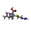  Mass: 301.362 Da / Num. of mol.: 1 / Source method: obtained synthetically / Formula: C12H19N3O4S / Comment: antibiotic*YM Mass: 301.362 Da / Num. of mol.: 1 / Source method: obtained synthetically / Formula: C12H19N3O4S / Comment: antibiotic*YM |
| #3: Chemical | ChemComp-GOL /   Mass: 92.094 Da / Num. of mol.: 1 / Source method: obtained synthetically / Formula: C3H8O3 Mass: 92.094 Da / Num. of mol.: 1 / Source method: obtained synthetically / Formula: C3H8O3 |
| #4: Water | ChemComp-HOH /  Mass: 18.015 Da / Num. of mol.: 369 / Source method: isolated from a natural source / Formula: H2O Mass: 18.015 Da / Num. of mol.: 369 / Source method: isolated from a natural source / Formula: H2O |
| Has protein modification | Y |
| Sequence details | AUTHORS STATE THAT THIS IS A SOLUBLE CONSTRUCT OF PBP 5. THE FIRST 29 AMINO ACIDS OF THE PROTEIN, ...AUTHORS STATE THAT THIS IS A SOLUBLE CONSTRUCT OF PBP 5. THE FIRST 29 AMINO ACIDS OF THE PROTEIN, WHICH ENCODE THE SIGNAL SEQUENCE, ARE NOT INCLUDED. THE LAST 17 AMINO ACIDS OF THE WILD-TYPE SEQUENCE ARE ALSO ABSENT AND ARE REPLACED BY SIX NON-NATIVE AMINO ACIDS (GDPVID - INTRODUCED |
-Experimental details
-Experiment
| Experiment | Method: X-RAY DIFFRACTION / Number of used crystals: 1 |
|---|
- Sample preparation
| Crystal | Density Matthews: 2.53 Å3/Da / Density % sol: 51.34 % |
|---|---|
| Crystal grow | Temperature: 294 K / Method: vapor diffusion, sitting drop / pH: 7 Details: 100 MM TRIS PH 7.0, 8 % PEG 400, VAPOR DIFFUSION, SITTING DROP, temperature 294K |
-Data collection
| Diffraction | Mean temperature: 100 K |
|---|---|
| Diffraction source | Source: SYNCHROTRON / Site: APS  / Beamline: 22-ID / Wavelength: 1 Å / Beamline: 22-ID / Wavelength: 1 Å |
| Detector | Type: MARMOSAIC 300 mm CCD / Detector: CCD / Date: Mar 24, 2004 |
| Radiation | Monochromator: DOUBLE CRYSTAL MONOCHROMATOR SI-220 / Protocol: SINGLE WAVELENGTH / Monochromatic (M) / Laue (L): M / Scattering type: x-ray |
| Radiation wavelength | Wavelength: 1 Å / Relative weight: 1 |
| Reflection | Resolution: 1.5→33 Å / Num. all: 61886 / Num. obs: 61886 / % possible obs: 96.6 % / Observed criterion σ(F): 0 / Observed criterion σ(I): 0 / Redundancy: 6.8 % / Biso Wilson estimate: 20.6 Å2 / Rmerge(I) obs: 0.055 / Rsym value: 0.055 / Net I/σ(I): 40.1 |
| Reflection shell | Resolution: 1.5→1.55 Å / Redundancy: 3.9 % / Rmerge(I) obs: 0.488 / Mean I/σ(I) obs: 1.8 / Num. unique all: 4880 / Rsym value: 0.488 / % possible all: 76.3 |
- Processing
| Software |
| |||||||||||||||||||||||||||||||||||||||||||||||||||||||||||||||||||||||||||||||||||||||||||||||||||||||||
|---|---|---|---|---|---|---|---|---|---|---|---|---|---|---|---|---|---|---|---|---|---|---|---|---|---|---|---|---|---|---|---|---|---|---|---|---|---|---|---|---|---|---|---|---|---|---|---|---|---|---|---|---|---|---|---|---|---|---|---|---|---|---|---|---|---|---|---|---|---|---|---|---|---|---|---|---|---|---|---|---|---|---|---|---|---|---|---|---|---|---|---|---|---|---|---|---|---|---|---|---|---|---|---|---|---|---|
| Refinement | Method to determine structure: FOURIER SYNTHESIS Starting model: PDB entry 1NZO Resolution: 1.5→32.97 Å / Cor.coef. Fo:Fc: 0.965 / Cor.coef. Fo:Fc free: 0.958 / SU B: 3.01 / SU ML: 0.051 / Isotropic thermal model: isotropic / Cross valid method: THROUGHOUT / σ(F): 0 / σ(I): 0 / ESU R Free: 0.077 / Stereochemistry target values: MAXIMUM LIKELIHOOD
| |||||||||||||||||||||||||||||||||||||||||||||||||||||||||||||||||||||||||||||||||||||||||||||||||||||||||
| Solvent computation | Ion probe radii: 0.8 Å / Shrinkage radii: 0.8 Å / VDW probe radii: 1.4 Å / Solvent model: MASK | |||||||||||||||||||||||||||||||||||||||||||||||||||||||||||||||||||||||||||||||||||||||||||||||||||||||||
| Displacement parameters | Biso mean: 22.92 Å2
| |||||||||||||||||||||||||||||||||||||||||||||||||||||||||||||||||||||||||||||||||||||||||||||||||||||||||
| Refinement step | Cycle: LAST / Resolution: 1.5→32.97 Å
| |||||||||||||||||||||||||||||||||||||||||||||||||||||||||||||||||||||||||||||||||||||||||||||||||||||||||
| Refine LS restraints |
| |||||||||||||||||||||||||||||||||||||||||||||||||||||||||||||||||||||||||||||||||||||||||||||||||||||||||
| LS refinement shell | Resolution: 1.5→1.539 Å / Total num. of bins used: 20
|
