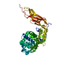
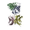
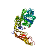
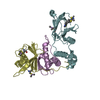
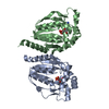
SEQUENCE TO PRODUCE SPBP 5, THE LAST 17 AMINO ACIDS WERE REMOVED BY DELETION OF THEIR RESPECTIVE ...SEQUENCE TO PRODUCE SPBP 5, THE LAST 17 AMINO ACIDS WERE REMOVED BY DELETION OF THEIR RESPECTIVE CODONS, AN ADDITIONAL SIX AMINO ACIDS (GDPVID) WERE INTRODUCED AT THE C TERMINUS DUE TO READ-THROUGH TO THE STOP CODON. NONE OF THESE NON-NATIVE RESIDUES ARE VISIBLE IN THE ELECTRON DENSITY MAP. THE FIRST 29 AMINO ACIDS OF THE PROTEIN ENCODED BY THE OPEN READING FRAME REPRESENT THE SIGNAL SEQUENCE, WHICH IS REMOVED DURING MATURATION AND TRANSPORT TO THE PERIPLASMIC SPACE AND IS NOT PRESENT IN THIS CONSTRUCT.
In the structure databanks used in Yorodumi, some data are registered as the other names, "COVID-19 virus" and "2019-nCoV". Here are the details of the virus and the list of structure data.
Jan 31, 2019. EMDB accession codes are about to change! (news from PDBe EMDB page)
EMDB accession codes are about to change! (news from PDBe EMDB page)
The allocation of 4 digits for EMDB accession codes will soon come to an end. Whilst these codes will remain in use, new EMDB accession codes will include an additional digit and will expand incrementally as the available range of codes is exhausted. The current 4-digit format prefixed with “EMD-” (i.e. EMD-XXXX) will advance to a 5-digit format (i.e. EMD-XXXXX), and so on. It is currently estimated that the 4-digit codes will be depleted around Spring 2019, at which point the 5-digit format will come into force.
The EM Navigator/Yorodumi systems omit the EMD- prefix.
Related info.:Q: What is EMD? / ID/Accession-code notation in Yorodumi/EM Navigator
Yorodumi is a browser for structure data from EMDB, PDB, SASBDB, etc.
This page is also the successor to EM Navigator detail page, and also detail information page/front-end page for Omokage search.
The word "yorodu" (or yorozu) is an old Japanese word meaning "ten thousand". "mi" (miru) is to see.
Related info.:EMDB / PDB / SASBDB / Comparison of 3 databanks / Yorodumi Search / Aug 31, 2016. New EM Navigator & Yorodumi / Yorodumi Papers / Jmol/JSmol / Function and homology information / Changes in new EM Navigator and Yorodumi
 Movie
Movie Controller

 Yorodumi
 Open data
 Basic information
 Components
 Keywords
 Function and homology information
 X-RAY DIFFRACTION /
  Authors
 Citation
 Structure visualization
 Downloads & links
 Other downloads


 PDBj
PDBj
 Assembly
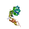
 Mass: 580.436 Da / Num. of mol.: 1 / Source method: obtained synthetically / Formula: C26H41BN4O10
 Mass: 92.094 Da / Num. of mol.: 1 / Source method: obtained synthetically / Formula: C3H8O3
 Mass: 18.015 Da / Num. of mol.: 351 / Source method: isolated from a natural source / Formula: H2O
 Sample preparation
 / Beamline: 22-ID / Wavelength: 1 Å
 Processing