 Movie
Movie Controller

[English] 日本語
 Yorodumi
- PDB-3mvg: Native structure of IRIP, a type I ribosome inactivating protein ... -
+ Open data

- Basic information
| Entry | Database: PDB / ID: 3mvg | ||||||
|---|---|---|---|---|---|---|---|
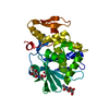
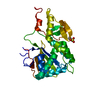
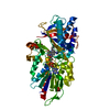
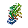
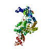
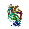
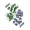
| Title | Native structure of IRIP, a type I ribosome inactivating protein from Iris hollandica var. at 1.25 A | ||||||
 Components Components | Ribosome inactivating type 1 protein | ||||||
 Keywords Keywords | HYDROLASE / Ribosome inactivating protein type I / IRIP / Pi-Loop | ||||||
| Function / homology |  Function and homology information Function and homology informationrRNA N-glycosylase / rRNA N-glycosylase activity / defense response / toxin activity / negative regulation of translation Similarity search - Function | ||||||
| Biological species |  Iris hollandica (Dutch iris) Iris hollandica (Dutch iris) | ||||||
| Method |  X-RAY DIFFRACTION / SYNCHROTRON / MOLECULAR REPLACEMENT / Resolution: 1.25 Å X-RAY DIFFRACTION / SYNCHROTRON / MOLECULAR REPLACEMENT / Resolution: 1.25 Å | ||||||
 Authors Authors | Meyer, A. / Weber, W. / Singh, T.P. / Betzel, C. | ||||||
 Citation Citation | Journal: to be published Title: Native structure of IRIP, a type I ribosome inactivating protein from Iris hollandica var. at 1.25 A Authors: Meyer, A. / Weber, W. / Singh, T.P. / Betzel, C. #1: Journal: Biochem.J. / Year: 1997 Title: Type 1 ribosome-inactivating proteins are the most abundant proteins in iris (Iris hollandica var. Professor Blaauw) bulbs: characterization and molecular cloning Authors: Van Damme, E.J. / Barre, A. / Barbieri, L. / Valbonesi, P. / Rouge, P. / Van Leuven, F. / Stirpe, F. / Peumans, W.J. #2: Journal: Protein Eng. / Year: 1992 Title: Analysis of several key active site residues of ricin A chain by mutagenesis and X-ray crystallography Authors: Kim, Y. / Robertus, J.D. #3: Journal: Proteins / Year: 1991 Title: Site-directed mutagenesis of ricin A-chain and implications for the mechanism of action Authors: Ready, M.P. / Kim, Y. / Robertus, J.D. | ||||||
| History |
|
- Structure visualization
| Structure viewer | Molecule: MolmilJmol/JSmol |
|---|
- Downloads & links
-Download
| PDBx/mmCIF format | 3mvg.cif.gz | 138.7 KB | Display | PDBx/mmCIF format |
|---|---|---|---|---|
| PDB format | pdb3mvg.ent.gz | 108.8 KB | Display | PDB format |
| PDBx/mmJSON format | 3mvg.json.gz | Tree view | PDBx/mmJSON format | |
| Others |  Other downloads Other downloads |
-Validation report
| Arichive directory | https://data.pdbj.org/pub/pdb/validation_reports/mv/3mvgftp://data.pdbj.org/pub/pdb/validation_reports/mv/3mvg | HTTPS FTP |
|---|
-Related structure data
| Related structure data |  1m2tS S: Starting model for refinement |
|---|---|
| Similar structure data |


-Links
 PDBj
PDBj

- Assembly
| Deposited unit | 
| ||||||||
|---|---|---|---|---|---|---|---|---|---|
| 1 |
| ||||||||
| Unit cell |
| ||||||||
| Details | THE IRIP DIMER IN THE ASYMMETRIC UNIT HAS BEEN FOUND IN AN NON-CRYSTALLOGRAPHIC 2-FOLD SYMMETRY. FORMATION OF THAT DIMER HAS PROBABLY NO BIOLOGICAL MEANING AND IS A RESULT OF THE CRYSTALLIZATION PROCESS. |
-Components
| #1: Protein | Mass: 31064.094 Da / Num. of mol.: 2 / Source method: isolated from a natural source / Source: (natural) Iris hollandica (Dutch iris) / References: UniProt: O04358, rRNA N-glycosylase#2: Chemical | ChemComp-SO4 /   Mass: 96.063 Da / Num. of mol.: 5 / Source method: obtained synthetically / Formula: SO4 Mass: 96.063 Da / Num. of mol.: 5 / Source method: obtained synthetically / Formula: SO4#3: Chemical | ChemComp-GOL /   Mass: 92.094 Da / Num. of mol.: 14 / Source method: obtained synthetically / Formula: C3H8O3 Mass: 92.094 Da / Num. of mol.: 14 / Source method: obtained synthetically / Formula: C3H8O3#4: Water | ChemComp-HOH / |  Mass: 18.015 Da / Num. of mol.: 756 / Source method: isolated from a natural source / Formula: H2O Mass: 18.015 Da / Num. of mol.: 756 / Source method: isolated from a natural source / Formula: H2OSequence details | THERE ARE CONFLICTS BETWEEN THE SEQUENCE AND DATABASE REFERENCE SEQUENCE. THE RESIDUES ARE ...THERE ARE CONFLICTS BETWEEN THE SEQUENCE AND DATABASE REFERENCE SEQUENCE. THE RESIDUES ARE CONSISTENT | |
|---|
-Experimental details
-Experiment
| Experiment | Method: X-RAY DIFFRACTION / Number of used crystals: 1 |
|---|
- Sample preparation
| Crystal | Density Matthews: 2.33 Å3/Da / Density % sol: 47.23 % |
|---|---|
| Crystal grow | Temperature: 293 K / Method: vapor diffusion, hanging drop / pH: 5 Details: 1.9M ammonium sulfate in 20% w/v glycerol, pH 5.0, VAPOR DIFFUSION, HANGING DROP, temperature 293K |
-Data collection
| Diffraction | Mean temperature: 100 K |
|---|---|
| Diffraction source | Source: SYNCHROTRON / Site: EMBL/DESY, HAMBURG  / Beamline: X11 / Wavelength: 0.8148 Å / Beamline: X11 / Wavelength: 0.8148 Å |
| Detector | Type: MAR CCD 165 mm / Detector: CCD / Date: Jul 3, 2007 |
| Radiation | Protocol: SINGLE WAVELENGTH / Monochromatic (M) / Laue (L): M / Scattering type: x-ray |
| Radiation wavelength | Wavelength: 0.8148 Å / Relative weight: 1 |
| Reflection | Resolution: 1.25→30 Å / Num. obs: 157596 / % possible obs: 99.8 % / Observed criterion σ(I): 2.6 / Redundancy: 4.4 % / Biso Wilson estimate: 17.62 Å2 / Rmerge(I) obs: 0.051 / Rsym value: 0.447 / Net I/σ(I): 25.6 / Num. measured all: 1839695 |
| Reflection shell | Resolution: 1.247→1.28 Å / Redundancy: 4.4 % / Rmerge(I) obs: 0.649 / Mean I/σ(I) obs: 2.67 / Num. unique all: 7666 / Rsym value: 0.45 / % possible all: 98 |
- Processing
| Software |
| |||||||||||||||||||||||||||||||||||||||||||||||||||||||||||||||||
|---|---|---|---|---|---|---|---|---|---|---|---|---|---|---|---|---|---|---|---|---|---|---|---|---|---|---|---|---|---|---|---|---|---|---|---|---|---|---|---|---|---|---|---|---|---|---|---|---|---|---|---|---|---|---|---|---|---|---|---|---|---|---|---|---|---|---|
| Refinement | Method to determine structure: MOLECULAR REPLACEMENT Starting model: PDB ENTRY 1M2T Resolution: 1.25→30 Å / Cor.coef. Fo:Fc: 0.974 / Cor.coef. Fo:Fc free: 0.969 / SU B: 0.646 / SU ML: 0.029 / Cross valid method: THROUGHOUT / ESU R Free: 0.044 / Stereochemistry target values: MAXIMUM LIKELIHOOD Details: HYDROGENS HAVE BEEN ADDED IN THE RIDING POSITIONS; Ala 93 (A/B) and Glycin 251 were refined based on the electron density results. Results are discussed in the publication.
| |||||||||||||||||||||||||||||||||||||||||||||||||||||||||||||||||
| Solvent computation | Ion probe radii: 0.8 Å / Shrinkage radii: 0.8 Å / VDW probe radii: 1.4 Å / Solvent model: MASK | |||||||||||||||||||||||||||||||||||||||||||||||||||||||||||||||||
| Displacement parameters | Biso mean: 17.883 Å2
| |||||||||||||||||||||||||||||||||||||||||||||||||||||||||||||||||
| Refinement step | Cycle: LAST / Resolution: 1.25→30 Å
| |||||||||||||||||||||||||||||||||||||||||||||||||||||||||||||||||
| Refine LS restraints |
| |||||||||||||||||||||||||||||||||||||||||||||||||||||||||||||||||
| LS refinement shell | Resolution: 1.247→1.28 Å / Total num. of bins used: 20
|
