 Movie
Movie Controller


+ Open data

- Basic information
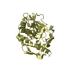
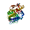
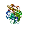
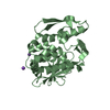
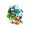
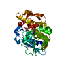
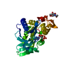
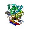
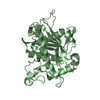
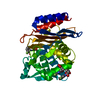
| Entry | Database: PDB / ID: 1bry | ||||||
|---|---|---|---|---|---|---|---|
| Title | BRYODIN TYPE I RIP | ||||||
 Components Components | BRYODIN I | ||||||
 Keywords Keywords | RIBOSOME-INACTIVATING PROTEIN / IMMUNOTOXIN / BRYODIN | ||||||
| Function / homology |  Function and homology information Function and homology informationrRNA N-glycosylase / rRNA N-glycosylase activity / defense response / toxin activity / negative regulation of translation Similarity search - Function | ||||||
| Biological species |  Bryonia dioica (red bryony) Bryonia dioica (red bryony) | ||||||
| Method |  X-RAY DIFFRACTION / MOLECULAR REPLACEMENT / Resolution: 2.1 Å X-RAY DIFFRACTION / MOLECULAR REPLACEMENT / Resolution: 2.1 Å | ||||||
 Authors Authors | Klei, H.E. / Chang, C.Y. | ||||||
 Citation Citation | Journal: Biochemistry / Year: 1997 Title: Molecular, biological, and preliminary structural analysis of recombinant bryodin 1, a ribosome-inactivating protein from the plant Bryonia dioica. Authors: Gawlak, S.L. / Neubauer, M. / Klei, H.E. / Chang, C.Y. / Einspahr, H.M. / Siegall, C.B. #1: Journal: Bioconjug.Chem. / Year: 1994Title: Characterization of Ribosome-Inactivating Proteins Isolated from Bryonia Dioica and Their Utility as Carcinoma-Reactive Immunoconjugates Authors: Siegall, C.B. / Gawlak, S.L. / Chace, D. / Wolff, E.A. / Mixan, B. / Marquardt, H. | ||||||
| History |
|
- Structure visualization
| Structure viewer | Molecule: MolmilJmol/JSmol |
|---|
- Downloads & links
-Download
| PDBx/mmCIF format | 1bry.cif.gz | 100.9 KB | Display | PDBx/mmCIF format |
|---|---|---|---|---|
| PDB format | pdb1bry.ent.gz | 79.1 KB | Display | PDB format |
| PDBx/mmJSON format | 1bry.json.gz | Tree view | PDBx/mmJSON format | |
| Others |  Other downloads Other downloads |
-Validation report
| Arichive directory | https://data.pdbj.org/pub/pdb/validation_reports/br/1bryftp://data.pdbj.org/pub/pdb/validation_reports/br/1bry | HTTPS FTP |
|---|
-Related structure data
| Related structure data |  1tcsS S: Starting model for refinement |
|---|---|
| Similar structure data |
-Links
 PDBj
PDBj

- Assembly
| Deposited unit | 
| ||||||||
|---|---|---|---|---|---|---|---|---|---|
| 1 | 
| ||||||||
| 2 | 
| ||||||||
| Unit cell |
| ||||||||
| Noncrystallographic symmetry (NCS) | NCS oper: (Code: given Matrix: (0.962022, -0.161317, -0.220205), Vector: |
-Components
| #1: Protein | Mass: 27202.820 Da / Num. of mol.: 2 / Fragment: RESIDUES 1 - 247 Source method: isolated from a genetically manipulated source Source: (gene. exp.) Bryonia dioica (red bryony)Description: CDNA SEQUENCE ISOLATED FROM BRYONIA DIOICA LEAF MRNA Organ: LEAF / Plasmid: PSE 13.0 / Production host:  |
|---|
-Experimental details
-Experiment
| Experiment | Method: X-RAY DIFFRACTION / Number of used crystals: 1 |
|---|
- Sample preparation
| Crystal | Density Matthews: 2.7 Å3/Da / Density % sol: 53 % Description: TRICHOSANTHIN MODEL TRUNCATED WITH PROGRAM MUTATE (S.SHERIFF) TO SELECT COMMON ATOMS BASED ON SEQUENCE ALIGNMENT. |
|---|---|
| Crystal grow | pH: 6 Details: 0.1M MES (PH 6.0), 23% MEPEG5K, 0.2M AMMONIUM SULFATE, 0.1M LICL |
| Crystal grow | *PLUS Method: unknown |
| Components of the solutions | *PLUS Common name: PEG5000 |
-Data collection
| Diffraction | Mean temperature: 100 K |
|---|---|
| Diffraction source | Source: ROTATING ANODE / Type: RIGAKU RUH2R / Wavelength: 1.5418 |
| Detector | Type: RIGAKU RAXIS II / Detector: IMAGE PLATE / Date: Jul 2, 1996 / Details: MIRRORS |
| Radiation | Monochromatic (M) / Laue (L): M / Scattering type: x-ray |
| Radiation wavelength | Wavelength: 1.5418 Å / Relative weight: 1 |
| Reflection | Resolution: 2.1→34 Å / Num. obs: 31074 / % possible obs: 89.1 % / Observed criterion σ(I): 0 / Redundancy: 3.6 % / Rmerge(I) obs: 0.043 / Net I/σ(I): 25.9 |
| Reflection shell | Resolution: 2.1→2.2 Å / Redundancy: 2.5 % / Rmerge(I) obs: 0.109 / Mean I/σ(I) obs: 8.7 / % possible all: 68.7 |
- Processing
| Software |
| ||||||||||||||||||||||||||||||||||||||||||||||||||||||||||||
|---|---|---|---|---|---|---|---|---|---|---|---|---|---|---|---|---|---|---|---|---|---|---|---|---|---|---|---|---|---|---|---|---|---|---|---|---|---|---|---|---|---|---|---|---|---|---|---|---|---|---|---|---|---|---|---|---|---|---|---|---|---|
| Refinement | Method to determine structure: MOLECULAR REPLACEMENT Starting model: PDB ENTRY 1TCS Resolution: 2.1→8 Å / σ(F): 1 Details: NCS RESTRAINTS BETWEEN THE TWO MOLECULES IN THE ASYMMETRIC UNIT WERE USED FOR THE FIRST TWO REFINEMENT CYCLES BUT WERE REMOVED FOR ALL SUBSEQUENT CYCLES WHEN RFREE WAS SHOWN TO DECREASE BY ...Details: NCS RESTRAINTS BETWEEN THE TWO MOLECULES IN THE ASYMMETRIC UNIT WERE USED FOR THE FIRST TWO REFINEMENT CYCLES BUT WERE REMOVED FOR ALL SUBSEQUENT CYCLES WHEN RFREE WAS SHOWN TO DECREASE BY APPROXIMATELY THE SAME AMOUNT AS R WHEN THE RESTRAINTS WERE REMOVED.
| ||||||||||||||||||||||||||||||||||||||||||||||||||||||||||||
| Displacement parameters | Biso mean: 13 Å2 | ||||||||||||||||||||||||||||||||||||||||||||||||||||||||||||
| Refinement step | Cycle: LAST / Resolution: 2.1→8 Å
| ||||||||||||||||||||||||||||||||||||||||||||||||||||||||||||
| Refine LS restraints |
| ||||||||||||||||||||||||||||||||||||||||||||||||||||||||||||
| Software | *PLUS Name: X-PLOR / Version: 3.1 / Classification: refinement | ||||||||||||||||||||||||||||||||||||||||||||||||||||||||||||
| Refine LS restraints | *PLUS
|
