 Movie
Movie Controller

[English] 日本語
 Yorodumi
- PDB-3mti: The Crystal Structure of a rRNA Methylase from Streptococcus ther... -
+ Open data

- Basic information
| Entry | Database: PDB / ID: 3mti | ||||||
|---|---|---|---|---|---|---|---|
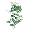
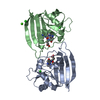
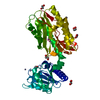
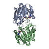
| Title | The Crystal Structure of a rRNA Methylase from Streptococcus thermophilus to 1.95A | ||||||
 Components Components | rRNA methylase | ||||||
 Keywords Keywords | TRANSFERASE / SAM-dependent / PSI / MCSG / Structural Genomics / Midwest Center for Structural Genomics / Protein Structure Initiative | ||||||
| Function / homology |  Function and homology information Function and homology information | ||||||
| Biological species |  Streptococcus thermophilus (bacteria) Streptococcus thermophilus (bacteria) | ||||||
| Method |  X-RAY DIFFRACTION / SYNCHROTRON / Resolution: 1.95 Å X-RAY DIFFRACTION / SYNCHROTRON / Resolution: 1.95 Å | ||||||
 Authors Authors | Stein, A.J. / Mulligan, R. / Freeman, L. / Joachimiak, A. / Midwest Center for Structural Genomics (MCSG) | ||||||
 Citation Citation | Journal: To be Published Title: The Crystal Structure of a rRNA Methylase from Streptococcus thermophilus to 1.95A Authors: Stein, A.J. / Mulligan, R. / Freeman, L. / Joachimiak, A. | ||||||
| History |
|
- Structure visualization
| Structure viewer | Molecule: MolmilJmol/JSmol |
|---|
- Downloads & links
-Download
| PDBx/mmCIF format | 3mti.cif.gz | 81.3 KB | Display | PDBx/mmCIF format |
|---|---|---|---|---|
| PDB format | pdb3mti.ent.gz | 61 KB | Display | PDB format |
| PDBx/mmJSON format | 3mti.json.gz | Tree view | PDBx/mmJSON format | |
| Others |  Other downloads Other downloads |
-Validation report
| Summary document | 3mti_validation.pdf.gz | 439.6 KB | Display | wwPDB validaton report |
|---|---|---|---|---|
| Full document | 3mti_full_validation.pdf.gz | 444.2 KB | Display | |
| Data in XML | 3mti_validation.xml.gz | 17.6 KB | Display | |
| Data in CIF | 3mti_validation.cif.gz | 23.1 KB | Display | |
| Arichive directory | https://data.pdbj.org/pub/pdb/validation_reports/mt/3mtiftp://data.pdbj.org/pub/pdb/validation_reports/mt/3mti | HTTPS FTP |
-Related structure data
| Similar structure data | |
|---|---|
| Other databases |


-Links
 PDBj
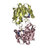
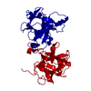
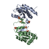
PDBj- Assembly
| Deposited unit | 
| ||||||||
|---|---|---|---|---|---|---|---|---|---|
| 1 |
| ||||||||
| Unit cell |
|
-Components
| #1: Protein | Mass: 20995.832 Da / Num. of mol.: 2 Source method: isolated from a genetically manipulated source Source: (gene. exp.) Streptococcus thermophilus (bacteria) / Strain: LMG 18311 / Gene: stu1645 / Plasmid: pMCSG7 / Production host: #2: Water | ChemComp-HOH / |  Mass: 18.015 Da / Num. of mol.: 125 / Source method: isolated from a natural source / Formula: H2O Mass: 18.015 Da / Num. of mol.: 125 / Source method: isolated from a natural source / Formula: H2OHas protein modification | Y | |
|---|
-Experimental details
-Experiment
| Experiment | Method: X-RAY DIFFRACTION / Number of used crystals: 1 |
|---|
- Sample preparation
| Crystal | Density Matthews: 2.41 Å3/Da / Density % sol: 49 % |
|---|---|
| Crystal grow | Temperature: 277 K / Method: vapor diffusion, sitting drop / pH: 7.5 Details: 25% PEG 3350, 0.1M HEPES pH 7.5, 0.2M Sodium acetate, VAPOR DIFFUSION, SITTING DROP, temperature 277K |
-Data collection
| Diffraction | Mean temperature: 100 K | ||||||||||||||||||||||||||||||||||||||||||||||||||||||||||||||||||
|---|---|---|---|---|---|---|---|---|---|---|---|---|---|---|---|---|---|---|---|---|---|---|---|---|---|---|---|---|---|---|---|---|---|---|---|---|---|---|---|---|---|---|---|---|---|---|---|---|---|---|---|---|---|---|---|---|---|---|---|---|---|---|---|---|---|---|---|
| Diffraction source | Source: SYNCHROTRON / Site: APS  / Beamline: 19-ID / Wavelength: 0.9794 Å / Beamline: 19-ID / Wavelength: 0.9794 Å | ||||||||||||||||||||||||||||||||||||||||||||||||||||||||||||||||||
| Detector | Type: ADSC QUANTUM 315 / Detector: CCD / Date: Jan 1, 2009 | ||||||||||||||||||||||||||||||||||||||||||||||||||||||||||||||||||
| Radiation | Monochromator: Double crystal / Protocol: SINGLE WAVELENGTH / Scattering type: x-ray | ||||||||||||||||||||||||||||||||||||||||||||||||||||||||||||||||||
| Radiation wavelength | Wavelength: 0.9794 Å / Relative weight: 1 | ||||||||||||||||||||||||||||||||||||||||||||||||||||||||||||||||||
| Reflection | Resolution: 1.95→50 Å / Num. obs: 28265 / % possible obs: 99.5 % / Redundancy: 3.3 % / Rmerge(I) obs: 0.085 / Χ2: 1.874 / Net I/σ(I): 8.1 | ||||||||||||||||||||||||||||||||||||||||||||||||||||||||||||||||||
| Reflection shell |
|
- Processing
| Software |
| |||||||||||||||||||||||||||||||||||||||||||||||||||||||||||||||||
|---|---|---|---|---|---|---|---|---|---|---|---|---|---|---|---|---|---|---|---|---|---|---|---|---|---|---|---|---|---|---|---|---|---|---|---|---|---|---|---|---|---|---|---|---|---|---|---|---|---|---|---|---|---|---|---|---|---|---|---|---|---|---|---|---|---|---|
| Refinement | Resolution: 1.95→50 Å / Cor.coef. Fo:Fc: 0.946 / Cor.coef. Fo:Fc free: 0.925 / SU B: 6.326 / SU ML: 0.168 / Cross valid method: THROUGHOUT / σ(F): 0 / ESU R Free: 0.175 / Stereochemistry target values: MAXIMUM LIKELIHOOD Details: HYDROGENS HAVE BEEN ADDED IN THE RIDING POSITIONS U VALUES : REFINED INDIVIDUALLY
| |||||||||||||||||||||||||||||||||||||||||||||||||||||||||||||||||
| Solvent computation | Ion probe radii: 0.8 Å / Shrinkage radii: 0.8 Å / VDW probe radii: 1.4 Å / Solvent model: MASK | |||||||||||||||||||||||||||||||||||||||||||||||||||||||||||||||||
| Displacement parameters | Biso mean: 36.071 Å2
| |||||||||||||||||||||||||||||||||||||||||||||||||||||||||||||||||
| Refinement step | Cycle: LAST / Resolution: 1.95→50 Å
| |||||||||||||||||||||||||||||||||||||||||||||||||||||||||||||||||
| Refine LS restraints |
| |||||||||||||||||||||||||||||||||||||||||||||||||||||||||||||||||
| LS refinement shell | Resolution: 1.949→1.999 Å / Total num. of bins used: 20
|
