 Movie
Movie Controller

[English] 日本語
 Yorodumi
- PDB-3moj: Structure of the RNA binding domain of the Bacillus subtilis YxiN... -
+ Open data

- Basic information
| Entry | Database: PDB / ID: 3moj | ||||||
|---|---|---|---|---|---|---|---|
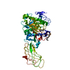
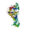
| Title | Structure of the RNA binding domain of the Bacillus subtilis YxiN protein complexed with a fragment of 23S ribosomal RNA | ||||||
 Components Components |
| ||||||
 Keywords Keywords | RNA binding protein/RNA / RNA / RNA recognition motif / RNA binding protein / ribosomal RNA / RNA binding protein-RNA complex | ||||||
| Function / homology |  Function and homology information Function and homology informationRNA strand annealing activity / 3'-5' RNA helicase activity / response to cold / ribosomal large subunit assembly / RNA helicase activity / RNA helicase / ATP hydrolysis activity / ATP binding / cytosol Similarity search - Function | ||||||
| Biological species |  | ||||||
| Method |  X-RAY DIFFRACTION / SYNCHROTRON / MOLECULAR REPLACEMENT / Resolution: 2.902 Å X-RAY DIFFRACTION / SYNCHROTRON / MOLECULAR REPLACEMENT / Resolution: 2.902 Å | ||||||
 Authors Authors | Hardin, J.W. / Hu, Y. / McKay, D.B. | ||||||
 Citation Citation | Journal: J.Mol.Biol. / Year: 2010 Title: Structure of the RNA binding domain of a DEAD-box helicase bound to its ribosomal RNA target reveals a novel mode of recognition by an RNA recognition motif. Authors: Hardin, J.W. / Hu, Y.X. / McKay, D.B. #1: Journal: Rna / Year: 2006Title: The domain of the Bacillus subtilis DEAD-box helicase YxiN that is responsible for specific binding of 23S rRNA has an RNA recognition motif fold. Authors: Wang, S. / Hu, Y. / Overgaard, M.T. / Karginov, F.V. / Uhlenbeck, O.C. / McKay, D.B. | ||||||
| History |
|
- Structure visualization
| Structure viewer | Molecule: MolmilJmol/JSmol |
|---|
- Downloads & links
-Download
| PDBx/mmCIF format | 3moj.cif.gz | 65.9 KB | Display | PDBx/mmCIF format |
|---|---|---|---|---|
| PDB format | pdb3moj.ent.gz | 46.2 KB | Display | PDB format |
| PDBx/mmJSON format | 3moj.json.gz | Tree view | PDBx/mmJSON format | |
| Others |  Other downloads Other downloads |
-Validation report
| Arichive directory | https://data.pdbj.org/pub/pdb/validation_reports/mo/3mojftp://data.pdbj.org/pub/pdb/validation_reports/mo/3moj | HTTPS FTP |
|---|
-Related structure data
| Similar structure data |
|---|


-Links
 PDBj
PDBj


- Assembly
| Deposited unit | 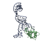
| ||||||||
|---|---|---|---|---|---|---|---|---|---|
| 1 |
| ||||||||
| Unit cell |
|
-Components
| #1: RNA chain | Mass: 23860.158 Da / Num. of mol.: 1 Fragment: E. coli 23 S ribosomal RNA (nucleotides 2508-2580 with GAAA tetraloop in nucleotides 2530-2533 and A2581 at 3' end) Source method: isolated from a genetically manipulated source Source: (gene. exp.) |
|---|---|
| #2: Protein | Mass: 8291.884 Da / Num. of mol.: 1 Source method: isolated from a genetically manipulated source Source: (gene. exp.) References: UniProt: P42305, Hydrolases; Acting on acid anhydrides; In phosphorus-containing anhydrides |
-Experimental details
-Experiment
| Experiment | Method: X-RAY DIFFRACTION / Number of used crystals: 1 |
|---|
- Sample preparation
| Crystal | Density Matthews: 3.7 Å3/Da / Density % sol: 66.73 % | ||||||||||||||||||||||||||||
|---|---|---|---|---|---|---|---|---|---|---|---|---|---|---|---|---|---|---|---|---|---|---|---|---|---|---|---|---|---|
| Crystal grow | Temperature: 290 K / Method: vapor diffusion / pH: 8 Details: 20 mg/ml protein-RNA complex with RNA in 5% molar excess, 5 mM Tris-HCL, 200 mM potassium acetate, 26-30% PEG-3350, pH 8.0, VAPOR DIFFUSION, temperature 290K | ||||||||||||||||||||||||||||
| Components of the solutions |
|
-Data collection
| Diffraction | Mean temperature: 100 K |
|---|---|
| Diffraction source | Source: SYNCHROTRON / Site: ALS  / Beamline: 8.2.1 / Wavelength: 1 Å / Beamline: 8.2.1 / Wavelength: 1 Å |
| Detector | Type: ADSC QUANTUM 315r / Detector: CCD / Date: Aug 1, 2009 |
| Radiation | Monochromator: Double crystal, Si(111) / Protocol: SINGLE WAVELENGTH / Monochromatic (M) / Laue (L): M / Scattering type: x-ray |
| Radiation wavelength | Wavelength: 1 Å / Relative weight: 1 |
| Reflection | Resolution: 2.9→50 Å / Num. all: 11090 / Num. obs: 10490 / % possible obs: 94.3 % / Observed criterion σ(F): 0 / Observed criterion σ(I): 0 / Redundancy: 4.2 % / Rsym value: 0.054 |
| Reflection shell | Resolution: 2.9→3 Å / Redundancy: 3.1 % / Rsym value: 0.296 / % possible all: 71.6 |
- Processing
| Software |
| ||||||||||||||||||||||||||||||||||||||||||||||||||||||||
|---|---|---|---|---|---|---|---|---|---|---|---|---|---|---|---|---|---|---|---|---|---|---|---|---|---|---|---|---|---|---|---|---|---|---|---|---|---|---|---|---|---|---|---|---|---|---|---|---|---|---|---|---|---|---|---|---|---|
| Refinement | Method to determine structure: MOLECULAR REPLACEMENT / Resolution: 2.902→19.896 Å / SU ML: 0.31 / σ(F): 0.05 / Stereochemistry target values: ML
| ||||||||||||||||||||||||||||||||||||||||||||||||||||||||
| Solvent computation | Shrinkage radii: 0.9 Å / VDW probe radii: 1.11 Å / Solvent model: FLAT BULK SOLVENT MODEL / Bsol: 50 Å2 / ksol: 0.23 e/Å3 | ||||||||||||||||||||||||||||||||||||||||||||||||||||||||
| Refinement step | Cycle: LAST / Resolution: 2.902→19.896 Å
| ||||||||||||||||||||||||||||||||||||||||||||||||||||||||
| Refine LS restraints |
| ||||||||||||||||||||||||||||||||||||||||||||||||||||||||
| LS refinement shell |
|
