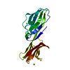
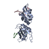
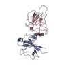
- PDB-3mgj: Crystal structure of the Saccharop_dh_N domain of MJ1480 protein ... -
+
Open data
ID or keywords:
Loading...
-
Basic information
Entry
Database: PDB / ID: 3mgj
Title
Crystal structure of the Saccharop_dh_N domain of MJ1480 protein from Methanococcus jannaschii. Northeast Structural Genomics Consortium Target MjR83a.
Resolution: 2.7→2.8 Å / Redundancy: 7.5 % / Rmerge(I) obs: 0.605 / Mean I/σ(I) obs: 3.8 / Num. unique all: 1053 / % possible all: 100
-
Processing
Software
Name
Version
Classification
HKL-2000
datacollection
SHELXDE
phasing
PHENIX
(phenix.refine: 1.5_2)
refinement
DENZO
datareduction
SCALEPACK
datascaling
Refinement
Method to determine structure: SAD / Resolution: 2.703→33.155 Å / SU ML: 0.38 / Cross valid method: THROUGHOUT / σ(F): 1.89 / Stereochemistry target values: ML
Rfactor
Num. reflection
% reflection
Selection details
Rfree
0.2549
480
4.6 %
RANDOM
Rwork
0.1992
-
-
-
obs
0.2018
10432
99.34 %
-
Solvent computation
Shrinkage radii: 0.9 Å / VDW probe radii: 1.11 Å / Solvent model: FLAT BULK SOLVENT MODEL / Bsol: 59.433 Å2 / ksol: 0.4 e/Å3
Refinement step
Cycle: LAST / Resolution: 2.703→33.155 Å
Protein
Nucleic acid
Ligand
Solvent
Total
Num. atoms
1459
0
0
31
1490
Refine LS restraints
Refine-ID
Type
Dev ideal
Number
X-RAY DIFFRACTION
f_bond_d
0.01
1483
X-RAY DIFFRACTION
f_angle_d
1.261
1998
X-RAY DIFFRACTION
f_dihedral_angle_d
20.069
577
X-RAY DIFFRACTION
f_chiral_restr
0.077
226
X-RAY DIFFRACTION
f_plane_restr
0.006
263
LS refinement shell
Resolution (Å)
Rfactor Rfree
Num. reflection Rfree
Rfactor Rwork
Num. reflection Rwork
Refine-ID
% reflection obs (%)
2.7031-3.094
0.3243
173
0.2458
3312
X-RAY DIFFRACTION
100
3.094-3.8971
0.2645
174
0.1918
3336
X-RAY DIFFRACTION
100
3.8971-33.157
0.2225
133
0.1893
3304
X-RAY DIFFRACTION
98
Refinement TLS params.
Refine-ID: X-RAY DIFFRACTION
ID
Method
L11 (°2)
L12 (°2)
L13 (°2)
L22 (°2)
L23 (°2)
L33 (°2)
S11 (Å °)
S12 (Å °)
S13 (Å °)
S21 (Å °)
S22 (Å °)
S23 (Å °)
S31 (Å °)
S32 (Å °)
S33 (Å °)
T11 (Å2)
T12 (Å2)
T13 (Å2)
T22 (Å2)
T23 (Å2)
T33 (Å2)
Origin x (Å)
Origin y (Å)
Origin z (Å)
1
refined
0.3804
-0.3014
-0.0302
0.6854
0.0335
1.5698
0.0848
-0.0242
0.0287
-0.1086
-0.136
0.1106
-0.0294
0.1496
0.0702
0.23
-0.0058
0.0057
0.2303
0.0224
0.2375
-4.8168
61.3411
40.8279
2
1.6108
0.2222
1.4992
1.6489
0.7598
1.611
0.0502
0.0371
-0.0842
-0.1405
-0.0871
0.239
-0.1126
-0.2178
0.0511
0.1886
0.022
-0.0028
0.2333
-0.0119
0.2495
Refinement TLS group
Selection details: chain B
+
About Yorodumi
-
News
-
Feb 9, 2022. New format data for meta-information of EMDB entries
New format data for meta-information of EMDB entries
Version 3 of the EMDB header file is now the official format.
The previous official version 1.9 will be removed from the archive.
In the structure databanks used in Yorodumi, some data are registered as the other names, "COVID-19 virus" and "2019-nCoV". Here are the details of the virus and the list of structure data.
Jan 31, 2019. EMDB accession codes are about to change! (news from PDBe EMDB page)
EMDB accession codes are about to change! (news from PDBe EMDB page)
The allocation of 4 digits for EMDB accession codes will soon come to an end. Whilst these codes will remain in use, new EMDB accession codes will include an additional digit and will expand incrementally as the available range of codes is exhausted. The current 4-digit format prefixed with “EMD-” (i.e. EMD-XXXX) will advance to a 5-digit format (i.e. EMD-XXXXX), and so on. It is currently estimated that the 4-digit codes will be depleted around Spring 2019, at which point the 5-digit format will come into force.
The EM Navigator/Yorodumi systems omit the EMD- prefix.
Related info.:Q: What is EMD? / ID/Accession-code notation in Yorodumi/EM Navigator
Yorodumi is a browser for structure data from EMDB, PDB, SASBDB, etc.
This page is also the successor to EM Navigator detail page, and also detail information page/front-end page for Omokage search.
The word "yorodu" (or yorozu) is an old Japanese word meaning "ten thousand". "mi" (miru) is to see.
Related info.:EMDB / PDB / SASBDB / Comparison of 3 databanks / Yorodumi Search / Aug 31, 2016. New EM Navigator & Yorodumi / Yorodumi Papers / Jmol/JSmol / Function and homology information / Changes in new EM Navigator and Yorodumi
 Movie
Movie Controller

 Yorodumi
 Open data
 Basic information
 Components
 Keywords
 Function and homology information
 Methanocaldococcus jannaschii (archaea)
 X-RAY DIFFRACTION /
  Authors
 Citation
 Structure visualization
 Downloads & links
 Other downloads


 PDBj
PDBj

 Assembly
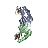

 Mass: 18.015 Da / Num. of mol.: 31 / Source method: isolated from a natural source / Formula: H2O
 Sample preparation
 / Beamline: X6A / Wavelength: 0.9791 Å
 Processing