 Movie
Movie Controller


+ Open data

- Basic information
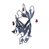
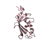
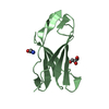
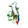
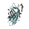
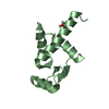
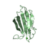
| Entry | Database: PDB / ID: 3fdo | ||||||
|---|---|---|---|---|---|---|---|
| Title | Structure of human MDMX in complex with high affinity peptide | ||||||
 Components Components |
| ||||||
 Keywords Keywords | CELL CYCLE / MDMX / MDM4 / MDM-X / MDM-4 / p53 / MDM2 | ||||||
| Function / homology |  Function and homology information Function and homology informationatrial septum development / heart valve development / atrioventricular valve morphogenesis / endocardial cushion morphogenesis / ventricular septum development / negative regulation of signal transduction by p53 class mediator / transcription repressor complex / DNA damage response, signal transduction by p53 class mediator / Stabilization of p53 / negative regulation of protein catabolic process ...atrial septum development / heart valve development / atrioventricular valve morphogenesis / endocardial cushion morphogenesis / ventricular septum development / negative regulation of signal transduction by p53 class mediator / transcription repressor complex / DNA damage response, signal transduction by p53 class mediator / Stabilization of p53 / negative regulation of protein catabolic process / Oncogene Induced Senescence / Regulation of TP53 Activity through Methylation / enzyme activator activity / ubiquitin-protein transferase activity / Regulation of TP53 Degradation / protein-containing complex assembly / Oxidative Stress Induced Senescence / cellular response to hypoxia / Regulation of TP53 Activity through Phosphorylation / regulation of cell cycle / protein stabilization / Ub-specific processing proteases / protein ubiquitination / negative regulation of cell population proliferation / negative regulation of DNA-templated transcription / negative regulation of apoptotic process / enzyme binding / negative regulation of transcription by RNA polymerase II / zinc ion binding / nucleoplasm / nucleus Similarity search - Function | ||||||
| Biological species |  Homo sapiens (human) Homo sapiens (human) | ||||||
| Method |  X-RAY DIFFRACTION / SYNCHROTRON / MOLECULAR REPLACEMENT / Resolution: 1.4 Å X-RAY DIFFRACTION / SYNCHROTRON / MOLECULAR REPLACEMENT / Resolution: 1.4 Å | ||||||
 Authors Authors | Czarna, A.L. / Popowicz, G.M. / Holak, T.A. | ||||||
 Citation Citation | Journal: Cell Cycle / Year: 2009 Title: High affinity interaction of the p53 peptide-analogue with human Mdm2 and Mdmx. Authors: Czarna, A. / Popowicz, G.M. / Pecak, A. / Wolf, S. / Dubin, G. / Holak, T.A. | ||||||
| History |
|
- Structure visualization
| Structure viewer | Molecule: MolmilJmol/JSmol |
|---|
- Downloads & links
-Download
| PDBx/mmCIF format | 3fdo.cif.gz | 61.7 KB | Display | PDBx/mmCIF format |
|---|---|---|---|---|
| PDB format | pdb3fdo.ent.gz | 44.4 KB | Display | PDB format |
| PDBx/mmJSON format | 3fdo.json.gz | Tree view | PDBx/mmJSON format | |
| Others |  Other downloads Other downloads |
-Validation report
| Arichive directory | https://data.pdbj.org/pub/pdb/validation_reports/fd/3fdoftp://data.pdbj.org/pub/pdb/validation_reports/fd/3fdo | HTTPS FTP |
|---|
-Related structure data
| Related structure data |  3g03C 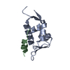 3dabS S: Starting model for refinement C: citing same article ( |
|---|---|
| Similar structure data |


-Links
 PDBj
PDBj


- Assembly
| Deposited unit | 
| ||||||||
|---|---|---|---|---|---|---|---|---|---|
| 1 | 
| ||||||||
| 2 | 
| ||||||||
| 3 | 
| ||||||||
| 4 | 
| ||||||||
| Unit cell |
|
-Components
| #1: Protein | Mass: 10274.058 Da / Num. of mol.: 1 / Fragment: p53 binding domain, UNP residues 23-111 Source method: isolated from a genetically manipulated source Source: (gene. exp.) Homo sapiens (human) / Gene: MDM4, MDMX / Plasmid: pET-46 / Production host:  | ||
|---|---|---|---|
| #2: Protein/peptide | Mass: 1496.641 Da / Num. of mol.: 1 / Source method: obtained synthetically / Details: Synthetic peptide designed to bind MDMX | ||
| #3: Chemical | ChemComp-MG /   Mass: 24.305 Da / Num. of mol.: 5 / Source method: obtained synthetically / Formula: Mg Mass: 24.305 Da / Num. of mol.: 5 / Source method: obtained synthetically / Formula: Mg#4: Water | ChemComp-HOH / |  Mass: 18.015 Da / Num. of mol.: 165 / Source method: isolated from a natural source / Formula: H2O Mass: 18.015 Da / Num. of mol.: 165 / Source method: isolated from a natural source / Formula: H2O |
-Experimental details
-Experiment
| Experiment | Method: X-RAY DIFFRACTION / Number of used crystals: 1 |
|---|
- Sample preparation
| Crystal | Density Matthews: 1.89 Å3/Da / Density % sol: 35.02 % |
|---|---|
| Crystal grow | Temperature: 290 K / Method: vapor diffusion, sitting drop / pH: 7.3 Details: 4.3M NaCl, 100mM Hepes, pH7.3, VAPOR DIFFUSION, SITTING DROP, temperature 290K |
-Data collection
| Diffraction | Mean temperature: 90 K |
|---|---|
| Diffraction source | Source: SYNCHROTRON / Site: SLS  / Beamline: X10SA / Wavelength: 1 Å / Beamline: X10SA / Wavelength: 1 Å |
| Detector | Type: MARRESEARCH / Detector: CCD / Date: Jul 15, 2008 / Details: monochromator |
| Radiation | Monochromator: GRAPHITE / Protocol: SINGLE WAVELENGTH / Monochromatic (M) / Laue (L): M / Scattering type: x-ray |
| Radiation wavelength | Wavelength: 1 Å / Relative weight: 1 |
| Reflection | Resolution: 1.4→30 Å / Num. all: 17646 / Num. obs: 14843 / % possible obs: 84.1 % / Observed criterion σ(F): 2 / Observed criterion σ(I): 2 |
| Reflection shell | Resolution: 1.4→1.5 Å / % possible all: 46.2 |
- Processing
| Software |
| ||||||||||||||||||||||||||||||||||||||||||||||||||||||||||||||||||||||||||||||||
|---|---|---|---|---|---|---|---|---|---|---|---|---|---|---|---|---|---|---|---|---|---|---|---|---|---|---|---|---|---|---|---|---|---|---|---|---|---|---|---|---|---|---|---|---|---|---|---|---|---|---|---|---|---|---|---|---|---|---|---|---|---|---|---|---|---|---|---|---|---|---|---|---|---|---|---|---|---|---|---|---|---|
| Refinement | Method to determine structure: MOLECULAR REPLACEMENT Starting model: PDB ENTRY 3DAB Resolution: 1.4→15 Å / Cor.coef. Fo:Fc: 0.962 / Cor.coef. Fo:Fc free: 0.915 / SU B: 2.973 / SU ML: 0.054 / Cross valid method: THROUGHOUT / σ(F): 2 / ESU R: 0.118 / ESU R Free: 0.099 / Stereochemistry target values: MAXIMUM LIKELIHOOD / Details: HYDROGENS HAVE BEEN ADDED IN THE RIDING POSITIONS
| ||||||||||||||||||||||||||||||||||||||||||||||||||||||||||||||||||||||||||||||||
| Solvent computation | Ion probe radii: 0.8 Å / Shrinkage radii: 0.8 Å / VDW probe radii: 1.2 Å / Solvent model: MASK | ||||||||||||||||||||||||||||||||||||||||||||||||||||||||||||||||||||||||||||||||
| Displacement parameters | Biso mean: 19.143 Å2
| ||||||||||||||||||||||||||||||||||||||||||||||||||||||||||||||||||||||||||||||||
| Refinement step | Cycle: LAST / Resolution: 1.4→15 Å
| ||||||||||||||||||||||||||||||||||||||||||||||||||||||||||||||||||||||||||||||||
| Refine LS restraints |
| ||||||||||||||||||||||||||||||||||||||||||||||||||||||||||||||||||||||||||||||||
| LS refinement shell | Resolution: 1.4→1.436 Å / Total num. of bins used: 20
|
