 Movie
Movie Controller

[English] 日本語
 Yorodumi
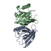
- PDB-3mep: Crystal Structure of ECA2234 protein from Erwinia carotovora, Nor... -
+ Open data

- Basic information
| Entry | Database: PDB / ID: 3mep | ||||||
|---|---|---|---|---|---|---|---|
| Title | Crystal Structure of ECA2234 protein from Erwinia carotovora, Northeast Structural Genomics Consortium Target EwR44 | ||||||
 Components Components | uncharacterized protein ECA2234 | ||||||
 Keywords Keywords | structural genomics / unknown function / all beta protein / PSI-2 / Protein Structure Initiative / Northeast Structural Genomics Consortium / NESG | ||||||
| Function / homology | Protein of unknown function DUF1349 / Uncharacterised conserved protein UCP022704 / Protein of unknown function (DUF1349) / Jelly Rolls - #200 / Concanavalin A-like lectin/glucanase domain superfamily / Jelly Rolls / Sandwich / Mainly Beta / Regulation of enolase protein 1 Function and homology information Function and homology information | ||||||
| Biological species |  Pectobacterium atrosepticum (bacteria) Pectobacterium atrosepticum (bacteria) | ||||||
| Method |  X-RAY DIFFRACTION / SYNCHROTRON / SAD / Resolution: 2.3 Å X-RAY DIFFRACTION / SYNCHROTRON / SAD / Resolution: 2.3 Å | ||||||
 Authors Authors | Seetharaman, J. / Abashidze, M. / Forouhar, F. / Mao, M. / Xiao, R. / Ciccosanti, C. / Wang, D. / Everett, J.K. / Nair, R. / Acton, T.B. ...Seetharaman, J. / Abashidze, M. / Forouhar, F. / Mao, M. / Xiao, R. / Ciccosanti, C. / Wang, D. / Everett, J.K. / Nair, R. / Acton, T.B. / Rost, B. / Montelione, G.T. / Tong, L. / Hunt, J.F. / Northeast Structural Genomics Consortium (NESG) | ||||||
 Citation Citation | Journal: To be Published Title: Northeast Structural Genomics Consortium Target EwR44 Authors: Seetharaman, J. / Abashidze, M. / Forouhar, F. / Mao, M. / Xiao, R. / Ciccosanti, C. / Wang, D. / Everett, J.K. / Nair, R. / Acton, T.B. / Rost, B. / Montelione, G.T. / Tong, L. / Hunt, J.F. | ||||||
| History |
|
- Structure visualization
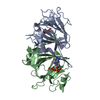
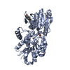
| Structure viewer | Molecule: MolmilJmol/JSmol |
|---|
- Downloads & links
-Download
| PDBx/mmCIF format | 3mep.cif.gz | 122.8 KB | Display | PDBx/mmCIF format |
|---|---|---|---|---|
| PDB format | pdb3mep.ent.gz | 96 KB | Display | PDB format |
| PDBx/mmJSON format | 3mep.json.gz | Tree view | PDBx/mmJSON format | |
| Others |  Other downloads Other downloads |
-Validation report
| Arichive directory | https://data.pdbj.org/pub/pdb/validation_reports/me/3mepftp://data.pdbj.org/pub/pdb/validation_reports/me/3mep | HTTPS FTP |
|---|
-Related structure data
| Similar structure data | |
|---|---|
| Other databases |


-Links
 PDBj
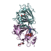
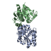
PDBj- Assembly
| Deposited unit | 
| ||||||||
|---|---|---|---|---|---|---|---|---|---|
| 1 | 
| ||||||||
| 2 | 
| ||||||||
| Unit cell |
|
-Components
| #1: Protein | Mass: 22757.896 Da / Num. of mol.: 3 Source method: isolated from a genetically manipulated source Source: (gene. exp.) Pectobacterium atrosepticum (bacteria) / Strain: SCRI1043 / Gene: ECA2234 / Plasmid: pET 21-23C / Production host: #2: Water | ChemComp-HOH / |  Mass: 18.015 Da / Num. of mol.: 293 / Source method: isolated from a natural source / Formula: H2O Mass: 18.015 Da / Num. of mol.: 293 / Source method: isolated from a natural source / Formula: H2OHas protein modification | Y | |
|---|
-Experimental details
-Experiment
| Experiment | Method: X-RAY DIFFRACTION / Number of used crystals: 1 |
|---|
- Sample preparation
| Crystal | Density Matthews: 2.2 Å3/Da / Density % sol: 44.14 % |
|---|---|
| Crystal grow | Temperature: 291 K / Method: vapor diffusion, hanging drop / pH: 7 Details: Protein solution: 100mM NaCl, 5mM DTT, 0.02% NaN3, 10mM Tris-HCl (pH 7.5) . Reservoir solution: 0.1M Hepes (pH 7), 16% PEG 8k, and 0.2M sodium nitrate., VAPOR DIFFUSION, HANGING DROP, temperature 291K |
-Data collection
| Diffraction | Mean temperature: 100 K |
|---|---|
| Diffraction source | Source: SYNCHROTRON / Site: NSLS  / Beamline: X4C / Wavelength: 0.97853 Å / Beamline: X4C / Wavelength: 0.97853 Å |
| Detector | Type: MAR CCD 165 mm / Detector: CCD / Date: Feb 11, 2010 / Details: mirrors |
| Radiation | Monochromator: Si 111 CHANNEL / Protocol: SINGLE WAVELENGTH / Monochromatic (M) / Laue (L): M / Scattering type: x-ray |
| Radiation wavelength | Wavelength: 0.97853 Å / Relative weight: 1 |
| Reflection | Resolution: 2.3→36.18 Å / Num. all: 53055 / Num. obs: 51199 / % possible obs: 96.5 % / Observed criterion σ(F): 0 / Observed criterion σ(I): 0 / Redundancy: 2.1 % / Biso Wilson estimate: 19.1 Å2 / Rmerge(I) obs: 0.082 / Rsym value: 0.069 / Net I/σ(I): 17.3 |
| Reflection shell | Resolution: 2.3→2.34 Å / Redundancy: 1.9 % / Rmerge(I) obs: 0.369 / Mean I/σ(I) obs: 3.05 / Rsym value: 0.3 / % possible all: 82.4 |
- Processing
| Software |
| ||||||||||||||||||||||||||||||||
|---|---|---|---|---|---|---|---|---|---|---|---|---|---|---|---|---|---|---|---|---|---|---|---|---|---|---|---|---|---|---|---|---|---|
| Refinement | Method to determine structure: SAD / Resolution: 2.3→36.18 Å / Rfactor Rfree error: 0.004 / Data cutoff high absF: 87013.344 / Data cutoff low absF: 0 / Isotropic thermal model: RESTRAINED / Cross valid method: THROUGHOUT / σ(F): 2 / σ(I): 2 / Stereochemistry target values: Engh & Huber
| ||||||||||||||||||||||||||||||||
| Solvent computation | Solvent model: FLAT MODEL / Bsol: 26.601 Å2 / ksol: 0.35 e/Å3 | ||||||||||||||||||||||||||||||||
| Displacement parameters | Biso mean: 31.4 Å2
| ||||||||||||||||||||||||||||||||
| Refine analyze |
| ||||||||||||||||||||||||||||||||
| Refinement step | Cycle: LAST / Resolution: 2.3→36.18 Å
| ||||||||||||||||||||||||||||||||
| Refine LS restraints |
| ||||||||||||||||||||||||||||||||
| LS refinement shell | Resolution: 2.3→2.44 Å / Rfactor Rfree error: 0.014 / Total num. of bins used: 6
| ||||||||||||||||||||||||||||||||
| Xplor file |
|
