 Movie
Movie Controller

[English] 日本語
 Yorodumi
- PDB-3mco: Crystal Structure of the 6-hyroxymethyl-7,8-dihydropterin pyropho... -
+ Open data

- Basic information
| Entry | Database: PDB / ID: 3mco | ||||||
|---|---|---|---|---|---|---|---|
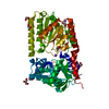
| Title | Crystal Structure of the 6-hyroxymethyl-7,8-dihydropterin pyrophosphokinase dihydropteroate synthase bifunctional enzyme from Francisella tularensis | ||||||
 Components Components | 2-amino-4-hydroxy-6-hydroxymethyldihydropteridine pyrophosphokinase/dihydropteroate synthase | ||||||
 Keywords Keywords | TRANSFERASE / folate / TIM barrel / kinase / synthase / HPPK / DHPS / pterin | ||||||
| Function / homology |  Function and homology information Function and homology information7,8-Dihydro-6-hydroxymethylpterin-pyrophosphokinase HPPK / Dihydropteroate synthase-like / Alpha-Beta Plaits / TIM Barrel / Alpha-Beta Barrel / 2-Layer Sandwich / Alpha Beta Similarity search - Domain/homology | ||||||
| Biological species |  Francisella tularensis subsp. holarctica (bacteria) Francisella tularensis subsp. holarctica (bacteria) | ||||||
| Method |  X-RAY DIFFRACTION / SYNCHROTRON / MOLECULAR REPLACEMENT / molecular replacement / Resolution: 2.3 Å X-RAY DIFFRACTION / SYNCHROTRON / MOLECULAR REPLACEMENT / molecular replacement / Resolution: 2.3 Å | ||||||
 Authors Authors | Pemble IV, C.W. / Mehta, P.K. / Mehra, S. / Li, Z. / Lee, R.E. / White, S.W. | ||||||
 Citation Citation | Journal: Plos One / Year: 2010 Title: Crystal structure of the 6-hydroxymethyl-7,8-dihydropterin pyrophosphokinase.dihydropteroate synthase bifunctional enzyme from Francisella tularensis. Authors: Pemble, C.W. / Mehta, P.K. / Mehra, S. / Li, Z. / Nourse, A. / Lee, R.E. / White, S.W. | ||||||
| History |
|
- Structure visualization
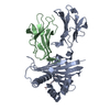
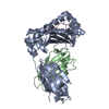
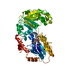
| Structure viewer | Molecule: MolmilJmol/JSmol |
|---|
- Downloads & links
-Download
| PDBx/mmCIF format | 3mco.cif.gz | 175.5 KB | Display | PDBx/mmCIF format |
|---|---|---|---|---|
| PDB format | pdb3mco.ent.gz | 135.9 KB | Display | PDB format |
| PDBx/mmJSON format | 3mco.json.gz | Tree view | PDBx/mmJSON format | |
| Others |  Other downloads Other downloads |
-Validation report
| Arichive directory | https://data.pdbj.org/pub/pdb/validation_reports/mc/3mcoftp://data.pdbj.org/pub/pdb/validation_reports/mc/3mco | HTTPS FTP |
|---|
-Related structure data
| Related structure data |  3mcmC  3mcnC  1twwS  2qx0S C: citing same article ( S: Starting model for refinement |
|---|---|
| Similar structure data |

-Links
 PDBj
PDBj


- Assembly
| Deposited unit | 
| ||||||||
|---|---|---|---|---|---|---|---|---|---|
| 1 | 
| ||||||||
| 2 | 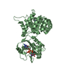
| ||||||||
| Unit cell |
|
-Components
| #1: Protein | Mass: 50587.254 Da / Num. of mol.: 2 Source method: isolated from a genetically manipulated source Source: (gene. exp.) Francisella tularensis subsp. holarctica (bacteria)Strain: LVS / Gene: folP/K, FTL_1265 / Plasmid: pET28 / Production host: #2: Chemical | ChemComp-PH2 /   Mass: 195.179 Da / Num. of mol.: 4 / Source method: obtained synthetically / Formula: C7H9N5O2 Mass: 195.179 Da / Num. of mol.: 4 / Source method: obtained synthetically / Formula: C7H9N5O2#3: Chemical |   Mass: 505.208 Da / Num. of mol.: 2 / Source method: obtained synthetically / Formula: C11H18N5O12P3 / Comment: AMP-CPP, energy-carrying molecule analogue*YM Mass: 505.208 Da / Num. of mol.: 2 / Source method: obtained synthetically / Formula: C11H18N5O12P3 / Comment: AMP-CPP, energy-carrying molecule analogue*YM#4: Chemical | ChemComp-MG /   Mass: 24.305 Da / Num. of mol.: 4 / Source method: obtained synthetically / Formula: Mg Mass: 24.305 Da / Num. of mol.: 4 / Source method: obtained synthetically / Formula: Mg#5: Water | ChemComp-HOH / |  Mass: 18.015 Da / Num. of mol.: 57 / Source method: isolated from a natural source / Formula: H2O Mass: 18.015 Da / Num. of mol.: 57 / Source method: isolated from a natural source / Formula: H2O |
|---|
-Experimental details
-Experiment
| Experiment | Method: X-RAY DIFFRACTION / Number of used crystals: 1 |
|---|
- Sample preparation
| Crystal | Density Matthews: 2.45 Å3/Da / Density % sol: 49.84 % |
|---|---|
| Crystal grow | Temperature: 291 K / Method: vapor diffusion, sitting drop / pH: 8 Details: TRIS, Na Acetate, PEG 4000, Glyerol, pH 8.0, vapor diffusion, sitting drop, temperature 291K |
-Data collection
| Diffraction | Mean temperature: 100 K |
|---|---|
| Diffraction source | Source: SYNCHROTRON / Site: APS  / Beamline: 22-ID / Wavelength: 1 Å / Beamline: 22-ID / Wavelength: 1 Å |
| Detector | Type: MARMOSAIC 300 mm CCD / Detector: CCD / Date: Dec 15, 2008 |
| Radiation | Protocol: SINGLE WAVELENGTH / Monochromatic (M) / Laue (L): M / Scattering type: x-ray |
| Radiation wavelength | Wavelength: 1 Å / Relative weight: 1 |
| Reflection | Resolution: 2.3→39.2 Å / Num. all: 42690 / Num. obs: 41121 / % possible obs: 96.3 % / Observed criterion σ(F): 0 / Observed criterion σ(I): 2 / Redundancy: 3.5 % / Rmerge(I) obs: 0.13 / Net I/σ(I): 24.8 |
| Reflection shell | Resolution: 2.3→2.378 Å / Redundancy: 2.8 % / Rmerge(I) obs: 0.3 / Mean I/σ(I) obs: 4.4 / Num. unique all: 3659 / % possible all: 84.9 |
-Phasing
| Phasing | Method: molecular replacement |
|---|
- Processing
| Software |
| |||||||||||||||||||||||||||||||||||||||||||||||||||||||||||||||||||||||||||||
|---|---|---|---|---|---|---|---|---|---|---|---|---|---|---|---|---|---|---|---|---|---|---|---|---|---|---|---|---|---|---|---|---|---|---|---|---|---|---|---|---|---|---|---|---|---|---|---|---|---|---|---|---|---|---|---|---|---|---|---|---|---|---|---|---|---|---|---|---|---|---|---|---|---|---|---|---|---|---|
| Refinement | Method to determine structure: MOLECULAR REPLACEMENT Starting model: PDB entries 2QX0, 1TWW Resolution: 2.3→38.251 Å / Occupancy max: 1 / Occupancy min: 1 / SU ML: 0.41 / Cross valid method: THROUGHOUT / σ(F): 1.98 / σ(I): 0 / Stereochemistry target values: ML
| |||||||||||||||||||||||||||||||||||||||||||||||||||||||||||||||||||||||||||||
| Solvent computation | Shrinkage radii: 0.9 Å / VDW probe radii: 1.11 Å / Solvent model: FLAT BULK SOLVENT MODEL / Bsol: 43.759 Å2 / ksol: 0.384 e/Å3 | |||||||||||||||||||||||||||||||||||||||||||||||||||||||||||||||||||||||||||||
| Displacement parameters | Biso max: 85.44 Å2 / Biso mean: 40.282 Å2 / Biso min: 14.1 Å2
| |||||||||||||||||||||||||||||||||||||||||||||||||||||||||||||||||||||||||||||
| Refinement step | Cycle: LAST / Resolution: 2.3→38.251 Å
| |||||||||||||||||||||||||||||||||||||||||||||||||||||||||||||||||||||||||||||
| Refine LS restraints |
| |||||||||||||||||||||||||||||||||||||||||||||||||||||||||||||||||||||||||||||
| LS refinement shell | Refine-ID: X-RAY DIFFRACTION / Total num. of bins used: 10
|
