 Movie
Movie Controller

[English] 日本語
 Yorodumi
- PDB-3mbo: Crystal Structure of the Glycosyltransferase BaBshA bound with UD... -
+ Open data

- Basic information
| Entry | Database: PDB / ID: 3mbo | ||||||
|---|---|---|---|---|---|---|---|
| Title | Crystal Structure of the Glycosyltransferase BaBshA bound with UDP and L-malate | ||||||
 Components Components | Glycosyltransferase, group 1 family | ||||||
 Keywords Keywords | TRANSFERASE / glycosyltransferase / bacillus anthracis / UDP / L-malate | ||||||
| Function / homology |  Function and homology information Function and homology informationbacillithiol biosynthetic process / Transferases; Glycosyltransferases; Hexosyltransferases / glycosyltransferase activity / nucleotide binding Similarity search - Function | ||||||
| Biological species |  | ||||||
| Method |  X-RAY DIFFRACTION / SYNCHROTRON / MOLECULAR REPLACEMENT / Resolution: 3.307 Å X-RAY DIFFRACTION / SYNCHROTRON / MOLECULAR REPLACEMENT / Resolution: 3.307 Å | ||||||
 Authors Authors | Wallace, B.D. / Claiborne, A. / Redinbo, M.R. | ||||||
 Citation Citation | Journal: Biochemistry / Year: 2010 Title: Characterization of the N-Acetyl-alpha-d-glucosaminyl l-Malate Synthase and Deacetylase Functions for Bacillithiol Biosynthesis in Bacillus anthracis Authors: Parsonage, D. / Newton, G.L. / Holder, R.C. / Wallace, B.D. / Paige, C. / Dos Santos, P.C. / Redinbo, M.R. / Fahey, R.C. / Reid, S.D. / Claiborne, A. | ||||||
| History |
|
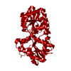
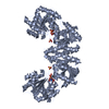
- Structure visualization
| Structure viewer | Molecule: MolmilJmol/JSmol |
|---|
- Downloads & links
-Download
| PDBx/mmCIF format | 3mbo.cif.gz | 595 KB | Display | PDBx/mmCIF format |
|---|---|---|---|---|
| PDB format | pdb3mbo.ent.gz | 485.2 KB | Display | PDB format |
| PDBx/mmJSON format | 3mbo.json.gz | Tree view | PDBx/mmJSON format | |
| Others |  Other downloads Other downloads |
-Validation report
| Arichive directory | https://data.pdbj.org/pub/pdb/validation_reports/mb/3mboftp://data.pdbj.org/pub/pdb/validation_reports/mb/3mbo | HTTPS FTP |
|---|
-Related structure data
| Related structure data | 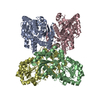 2jjmS S: Starting model for refinement |
|---|---|
| Similar structure data |


-Links
 PDBj
PDBj


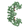
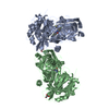
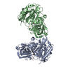
- Assembly
| Deposited unit | 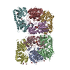
| ||||||||
|---|---|---|---|---|---|---|---|---|---|
| 1 | 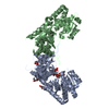
| ||||||||
| 2 | 
| ||||||||
| 3 | 
| ||||||||
| 4 | 
| ||||||||
| Unit cell |
| ||||||||
| Details | GEL FILTRATION EXPERIMENT SHOWS THAT THE BIOLOGICAL UNIT IS DIMER. |
-Components
-Protein , 1 types, 8 molecules ABCDEFGH
| #1: Protein | Mass: 46362.086 Da / Num. of mol.: 8 Source method: isolated from a genetically manipulated source Source: (gene. exp.) |
|---|
-Non-polymers , 5 types, 319 molecules 


| #2: Chemical | ChemComp-UDP /  Type: RNA linking / Mass: 404.161 Da / Num. of mol.: 4 / Source method: obtained synthetically / Formula: C9H14N2O12P2 / Comment: UDP*YM Type: RNA linking / Mass: 404.161 Da / Num. of mol.: 4 / Source method: obtained synthetically / Formula: C9H14N2O12P2 / Comment: UDP*YM#3: Chemical | ChemComp-GOL /  Mass: 92.094 Da / Num. of mol.: 21 / Source method: obtained synthetically / Formula: C3H8O3 Mass: 92.094 Da / Num. of mol.: 21 / Source method: obtained synthetically / Formula: C3H8O3#4: Chemical | ChemComp-MLT /  Mass: 134.087 Da / Num. of mol.: 4 / Source method: obtained synthetically / Formula: C4H6O5 Mass: 134.087 Da / Num. of mol.: 4 / Source method: obtained synthetically / Formula: C4H6O5#5: Chemical |  Mass: 24.305 Da / Num. of mol.: 2 / Source method: obtained synthetically / Formula: Mg Mass: 24.305 Da / Num. of mol.: 2 / Source method: obtained synthetically / Formula: Mg#6: Water | ChemComp-HOH / | Mass: 18.015 Da / Num. of mol.: 288 / Source method: isolated from a natural source / Formula: H2O |
|---|
-Details
| Has protein modification | Y |
|---|
-Experimental details
-Experiment
| Experiment | Method: X-RAY DIFFRACTION / Number of used crystals: 1 |
|---|
- Sample preparation
| Crystal | Density Matthews: 2.81 Å3/Da / Density % sol: 56.23 % |
|---|---|
| Crystal grow | Temperature: 289 K / Method: vapor diffusion, hanging drop / pH: 7.5 Details: 15% PEG 3350, 0.2 M MgFormate, 0.02% sodium azide, 1 mM L-malate, 1 mM Uridine diphosphate, pH 7.5, vapor diffusion, hanging drop, temperature 289K |
-Data collection
| Diffraction | Mean temperature: 100 K | ||||||||||||||||||||||||||||||||||||||||||||||||||||||||||||||||||||||||||||||||||||
|---|---|---|---|---|---|---|---|---|---|---|---|---|---|---|---|---|---|---|---|---|---|---|---|---|---|---|---|---|---|---|---|---|---|---|---|---|---|---|---|---|---|---|---|---|---|---|---|---|---|---|---|---|---|---|---|---|---|---|---|---|---|---|---|---|---|---|---|---|---|---|---|---|---|---|---|---|---|---|---|---|---|---|---|---|---|
| Diffraction source | Source: SYNCHROTRON / Site: APS  / Beamline: 22-BM / Wavelength: 1 Å / Beamline: 22-BM / Wavelength: 1 Å | ||||||||||||||||||||||||||||||||||||||||||||||||||||||||||||||||||||||||||||||||||||
| Detector | Type: MAR CCD 165 mm / Detector: CCD / Date: Jun 26, 2009 | ||||||||||||||||||||||||||||||||||||||||||||||||||||||||||||||||||||||||||||||||||||
| Radiation | Protocol: SINGLE WAVELENGTH / Monochromatic (M) / Laue (L): M / Scattering type: x-ray | ||||||||||||||||||||||||||||||||||||||||||||||||||||||||||||||||||||||||||||||||||||
| Radiation wavelength | Wavelength: 1 Å / Relative weight: 1 | ||||||||||||||||||||||||||||||||||||||||||||||||||||||||||||||||||||||||||||||||||||
| Reflection | Resolution: 3.3→50 Å / Num. obs: 57310 / % possible obs: 99.3 % / Redundancy: 5.7 % / Rmerge(I) obs: 0.169 / Net I/σ(I): 7.1 | ||||||||||||||||||||||||||||||||||||||||||||||||||||||||||||||||||||||||||||||||||||
| Reflection shell |
|
- Processing
| Software |
| |||||||||||||||||||||||||||||||||||||||||||||||||||||||||||||||||||||||||||||||||||||||||||||||||||||||||||||||||||||||||||||||||||||||||||||||||||
|---|---|---|---|---|---|---|---|---|---|---|---|---|---|---|---|---|---|---|---|---|---|---|---|---|---|---|---|---|---|---|---|---|---|---|---|---|---|---|---|---|---|---|---|---|---|---|---|---|---|---|---|---|---|---|---|---|---|---|---|---|---|---|---|---|---|---|---|---|---|---|---|---|---|---|---|---|---|---|---|---|---|---|---|---|---|---|---|---|---|---|---|---|---|---|---|---|---|---|---|---|---|---|---|---|---|---|---|---|---|---|---|---|---|---|---|---|---|---|---|---|---|---|---|---|---|---|---|---|---|---|---|---|---|---|---|---|---|---|---|---|---|---|---|---|---|---|---|---|
| Refinement | Method to determine structure: MOLECULAR REPLACEMENT Starting model: PDB ENTRY 2JJM Resolution: 3.307→48.222 Å / Occupancy max: 1 / Occupancy min: 0.75 / SU ML: 3.28 / σ(F): 0.08 / Phase error: 24.42 / Stereochemistry target values: ML
| |||||||||||||||||||||||||||||||||||||||||||||||||||||||||||||||||||||||||||||||||||||||||||||||||||||||||||||||||||||||||||||||||||||||||||||||||||
| Solvent computation | Shrinkage radii: 0.9 Å / VDW probe radii: 1.11 Å / Solvent model: FLAT BULK SOLVENT MODEL / Bsol: 41.359 Å2 / ksol: 0.307 e/Å3 | |||||||||||||||||||||||||||||||||||||||||||||||||||||||||||||||||||||||||||||||||||||||||||||||||||||||||||||||||||||||||||||||||||||||||||||||||||
| Displacement parameters | Biso mean: 74.394 Å2
| |||||||||||||||||||||||||||||||||||||||||||||||||||||||||||||||||||||||||||||||||||||||||||||||||||||||||||||||||||||||||||||||||||||||||||||||||||
| Refinement step | Cycle: LAST / Resolution: 3.307→48.222 Å
| |||||||||||||||||||||||||||||||||||||||||||||||||||||||||||||||||||||||||||||||||||||||||||||||||||||||||||||||||||||||||||||||||||||||||||||||||||
| Refine LS restraints |
| |||||||||||||||||||||||||||||||||||||||||||||||||||||||||||||||||||||||||||||||||||||||||||||||||||||||||||||||||||||||||||||||||||||||||||||||||||
| LS refinement shell |
|
