 Movie
Movie Controller

[English] 日本語
 Yorodumi
- PDB-3mac: crystal structure of GP41-derived protein complexed with fab 8062 -
+ Open data

- Basic information
| Entry | Database: PDB / ID: 3mac | ||||||
|---|---|---|---|---|---|---|---|
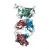
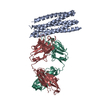
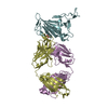
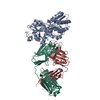
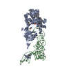
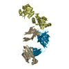
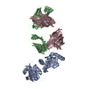
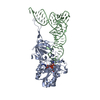
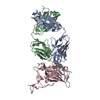
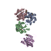
| Title | crystal structure of GP41-derived protein complexed with fab 8062 | ||||||
 Components Components |
| ||||||
 Keywords Keywords | IMMUNE SYSTEM / GP41 / fab8062 | ||||||
| Function / homology | Methane Monooxygenase Hydroxylase; Chain G, domain 1 - #1860 / Methane Monooxygenase Hydroxylase; Chain G, domain 1 / Immunoglobulins / Up-down Bundle / Immunoglobulin-like / Sandwich / Mainly Beta / Mainly Alpha / Transmembrane glycoprotein Function and homology information Function and homology information | ||||||
| Biological species |   Human immunodeficiency virus 1 Human immunodeficiency virus 1 Homo sapiens (human) Homo sapiens (human) | ||||||
| Method |  X-RAY DIFFRACTION / SYNCHROTRON / MOLECULAR REPLACEMENT / Resolution: 2.5 Å X-RAY DIFFRACTION / SYNCHROTRON / MOLECULAR REPLACEMENT / Resolution: 2.5 Å | ||||||
 Authors Authors | Li, M. / Gustchina, E. / Louis, J. / Gustchina, A. / Wlodawer, A. / Clore, M. | ||||||
 Citation Citation | Journal: Plos Pathog. / Year: 2010 Title: Structural Basis of HIV-1 Neutralization by Affinity Matured Fabs Directed against the Internal Trimeric Coiled-Coil of gp41. Authors: Gustchina, E. / Li, M. / Louis, J.M. / Anderson, D.E. / Lloyd, J. / Frisch, C. / Bewley, C.A. / Gustchina, A. / Wlodawer, A. / Clore, G.M. | ||||||
| History |
|
- Structure visualization
| Structure viewer | Molecule: MolmilJmol/JSmol |
|---|
- Downloads & links
-Download
| PDBx/mmCIF format | 3mac.cif.gz | 252 KB | Display | PDBx/mmCIF format |
|---|---|---|---|---|
| PDB format | pdb3mac.ent.gz | 203.9 KB | Display | PDB format |
| PDBx/mmJSON format | 3mac.json.gz | Tree view | PDBx/mmJSON format | |
| Others |  Other downloads Other downloads |
-Validation report
| Arichive directory | https://data.pdbj.org/pub/pdb/validation_reports/ma/3macftp://data.pdbj.org/pub/pdb/validation_reports/ma/3mac | HTTPS FTP |
|---|
-Related structure data
| Related structure data |  3ma9C  2cmrS C: citing same article ( S: Starting model for refinement |
|---|---|
| Similar structure data |
-Links
 PDBj
PDBj

- Assembly
| Deposited unit | 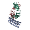
| ||||||||
|---|---|---|---|---|---|---|---|---|---|
| 1 |
| ||||||||
| Unit cell |
|
-Components
| #1: Protein | Mass: 24479.418 Da / Num. of mol.: 1 Source method: isolated from a genetically manipulated source Source: (gene. exp.) Human immunodeficiency virus 1 / Production host:  |
|---|---|
| #2: Antibody | Mass: 26334.322 Da / Num. of mol.: 1 Source method: isolated from a genetically manipulated source Source: (gene. exp.) Homo sapiens (human) |
| #3: Antibody | Mass: 22640.844 Da / Num. of mol.: 1 Source method: isolated from a genetically manipulated source Source: (gene. exp.) Homo sapiens (human) |
| #4: Water | ChemComp-HOH /  Mass: 18.015 Da / Num. of mol.: 69 / Source method: isolated from a natural source / Formula: H2O Mass: 18.015 Da / Num. of mol.: 69 / Source method: isolated from a natural source / Formula: H2O |
| Has protein modification | Y |
-Experimental details
-Experiment
| Experiment | Method: X-RAY DIFFRACTION / Number of used crystals: 1 |
|---|
- Sample preparation
| Crystal | Density Matthews: 2.34 Å3/Da / Density % sol: 47.36 % |
|---|---|
| Crystal grow | Temperature: 293 K / Method: vapor diffusion, hanging drop / pH: 8 Details: 14% Polyethylene glycol 10K, 0.1M ammonium sulfate, 5% ethylene glycol, pH 8.0, VAPOR DIFFUSION, HANGING DROP, temperature 293K |
-Data collection
| Diffraction | Mean temperature: 100 K |
|---|---|
| Diffraction source | Source: SYNCHROTRON / Site: APS  / Beamline: 22-ID / Wavelength: 1 Å / Beamline: 22-ID / Wavelength: 1 Å |
| Detector | Type: MARCCD300 / Detector: CCD / Date: Jan 1, 2009 / Details: mirror |
| Radiation | Monochromator: mirror / Protocol: SINGLE WAVELENGTH / Monochromatic (M) / Laue (L): M / Scattering type: x-ray |
| Radiation wavelength | Wavelength: 1 Å / Relative weight: 1 |
| Reflection | Resolution: 2.5→29.5 Å / Num. obs: 23655 / % possible obs: 99.1 % / Observed criterion σ(I): -3 / Redundancy: 3.1 % / Rsym value: 0.082 / Net I/σ(I): 13.79 |
| Reflection shell | Resolution: 2.5→2.59 Å / Redundancy: 3 % / Mean I/σ(I) obs: 2 / Rsym value: 0.524 / % possible all: 96.7 |
- Processing
| Software |
| ||||||||||||||||||||||||||||||||||||||||||||||||||||||||||||||||||||||||||||||||||||||||||||||||||||||||||||||||||||||||||||||||||||||||||||||||||||||||||||||||||||||||||
|---|---|---|---|---|---|---|---|---|---|---|---|---|---|---|---|---|---|---|---|---|---|---|---|---|---|---|---|---|---|---|---|---|---|---|---|---|---|---|---|---|---|---|---|---|---|---|---|---|---|---|---|---|---|---|---|---|---|---|---|---|---|---|---|---|---|---|---|---|---|---|---|---|---|---|---|---|---|---|---|---|---|---|---|---|---|---|---|---|---|---|---|---|---|---|---|---|---|---|---|---|---|---|---|---|---|---|---|---|---|---|---|---|---|---|---|---|---|---|---|---|---|---|---|---|---|---|---|---|---|---|---|---|---|---|---|---|---|---|---|---|---|---|---|---|---|---|---|---|---|---|---|---|---|---|---|---|---|---|---|---|---|---|---|---|---|---|---|---|---|---|---|
| Refinement | Method to determine structure: MOLECULAR REPLACEMENT Starting model: PDB ENTRY 2CMR Resolution: 2.5→29.5 Å / Cor.coef. Fo:Fc: 0.948 / Cor.coef. Fo:Fc free: 0.923 / SU B: 21.692 / SU ML: 0.224 / Cross valid method: THROUGHOUT / ESU R: 0.602 / ESU R Free: 0.31 / Stereochemistry target values: MAXIMUM LIKELIHOOD / Details: HYDROGENS HAVE BEEN ADDED IN THE RIDING POSITIONS
| ||||||||||||||||||||||||||||||||||||||||||||||||||||||||||||||||||||||||||||||||||||||||||||||||||||||||||||||||||||||||||||||||||||||||||||||||||||||||||||||||||||||||||
| Solvent computation | Ion probe radii: 0.8 Å / Shrinkage radii: 0.8 Å / VDW probe radii: 1.2 Å / Solvent model: MASK | ||||||||||||||||||||||||||||||||||||||||||||||||||||||||||||||||||||||||||||||||||||||||||||||||||||||||||||||||||||||||||||||||||||||||||||||||||||||||||||||||||||||||||
| Displacement parameters | Biso mean: 32.229 Å2
| ||||||||||||||||||||||||||||||||||||||||||||||||||||||||||||||||||||||||||||||||||||||||||||||||||||||||||||||||||||||||||||||||||||||||||||||||||||||||||||||||||||||||||
| Refinement step | Cycle: LAST / Resolution: 2.5→29.5 Å
| ||||||||||||||||||||||||||||||||||||||||||||||||||||||||||||||||||||||||||||||||||||||||||||||||||||||||||||||||||||||||||||||||||||||||||||||||||||||||||||||||||||||||||
| Refine LS restraints |
| ||||||||||||||||||||||||||||||||||||||||||||||||||||||||||||||||||||||||||||||||||||||||||||||||||||||||||||||||||||||||||||||||||||||||||||||||||||||||||||||||||||||||||
| LS refinement shell | Resolution: 2.502→2.567 Å / Total num. of bins used: 20
| ||||||||||||||||||||||||||||||||||||||||||||||||||||||||||||||||||||||||||||||||||||||||||||||||||||||||||||||||||||||||||||||||||||||||||||||||||||||||||||||||||||||||||
| Refinement TLS params. | Method: refined / Refine-ID: X-RAY DIFFRACTION
| ||||||||||||||||||||||||||||||||||||||||||||||||||||||||||||||||||||||||||||||||||||||||||||||||||||||||||||||||||||||||||||||||||||||||||||||||||||||||||||||||||||||||||
| Refinement TLS group |
|
