 Movie
Movie Controller


+ Open data

- Basic information
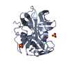
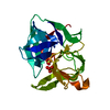
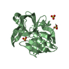
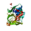
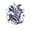
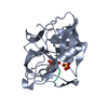
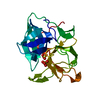
| Entry | Database: PDB / ID: 3m7t | ||||||
|---|---|---|---|---|---|---|---|
| Title | Crystal Structure of Alpha-Lytic Protease SB2+3 E8A/R105S Mutant | ||||||
 Components Components | Alpha-lytic protease | ||||||
 Keywords Keywords | HYDROLASE / Disulfide bond / Protease / Serine protease / Zymogen | ||||||
| Function / homology |  Function and homology information Function and homology informationalpha-lytic endopeptidase / serine-type endopeptidase activity / proteolysis / extracellular region Similarity search - Function | ||||||
| Biological species |  Lysobacter enzymogenes (bacteria) Lysobacter enzymogenes (bacteria) | ||||||
| Method |  X-RAY DIFFRACTION / SYNCHROTRON / MOLECULAR REPLACEMENT / Resolution: 1.55 Å X-RAY DIFFRACTION / SYNCHROTRON / MOLECULAR REPLACEMENT / Resolution: 1.55 Å | ||||||
 Authors Authors | Agard, D.A. / Erciyas Bailey, F.P. / Waddling, C.A. | ||||||
 Citation Citation | Journal: To be Published Title: Quantifying protein unfolding cooperativity with acid sensitive probes: Interdomain salt bridge contributions to unfolding cooperativity are combined efficiently in alpha-Lytic Protease Authors: Erciyas Bailey, F.P. / Waddling, C.A. / Agard, D.A. #1: Journal: J.Mol.Biol. / Year: 2004Title: The 0.83 A resolution crystal structure of alpha-lytic protease reveals the detailed structure of the active site and identifies a source of conformational strain. Authors: Fuhrmann, C.N. / Kelch, B.A. / Ota, N. / Agard, D.A. #2: Journal: Protein Sci. / Year: 1997Title: Conformational substates in enzyme mechanism: the 120 K structure of alpha-lytic protease at 1.5 A resolution. Authors: Rader, S.D. / Agard, D.A. | ||||||
| History |
|
- Structure visualization
| Structure viewer | Molecule: MolmilJmol/JSmol |
|---|
- Downloads & links
-Download
| PDBx/mmCIF format | 3m7t.cif.gz | 102 KB | Display | PDBx/mmCIF format |
|---|---|---|---|---|
| PDB format | pdb3m7t.ent.gz | 76.9 KB | Display | PDB format |
| PDBx/mmJSON format | 3m7t.json.gz | Tree view | PDBx/mmJSON format | |
| Others |  Other downloads Other downloads |
-Validation report
| Arichive directory | https://data.pdbj.org/pub/pdb/validation_reports/m7/3m7tftp://data.pdbj.org/pub/pdb/validation_reports/m7/3m7t | HTTPS FTP |
|---|
-Related structure data
| Related structure data |  3m7uC  1ssxS C: citing same article ( S: Starting model for refinement |
|---|---|
| Similar structure data |


-Links
 PDBj
PDBj- Assembly
| Deposited unit | 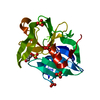
| |||||||||
|---|---|---|---|---|---|---|---|---|---|---|
| 1 |
| |||||||||
| Unit cell |
| |||||||||
| Components on special symmetry positions |
|
-Components
| #1: Protein | Mass: 19746.979 Da / Num. of mol.: 1 / Fragment: MATURE PROTEASE DOMAIN (RESIDUES 1-198) / Mutation: E8A, R105S Source method: isolated from a genetically manipulated source Source: (gene. exp.) Lysobacter enzymogenes (bacteria) / Gene: alpha-LP, Lysobacter / Plasmid: PALP12 / Production host: | ||||
|---|---|---|---|---|---|
| #2: Chemical | ChemComp-GOL /   Mass: 92.094 Da / Num. of mol.: 1 / Source method: obtained synthetically / Formula: C3H8O3 Mass: 92.094 Da / Num. of mol.: 1 / Source method: obtained synthetically / Formula: C3H8O3 | ||||
| #3: Chemical | ChemComp-SO4 /   Mass: 96.063 Da / Num. of mol.: 5 / Source method: obtained synthetically / Formula: SO4 Mass: 96.063 Da / Num. of mol.: 5 / Source method: obtained synthetically / Formula: SO4#4: Water | ChemComp-HOH / |  Mass: 18.015 Da / Num. of mol.: 448 / Source method: isolated from a natural source / Formula: H2O Mass: 18.015 Da / Num. of mol.: 448 / Source method: isolated from a natural source / Formula: H2OHas protein modification | Y | |
-Experimental details
-Experiment
| Experiment | Method: X-RAY DIFFRACTION / Number of used crystals: 1 |
|---|
- Sample preparation
| Crystal | Density Matthews: 2.54 Å3/Da / Density % sol: 51.58 % |
|---|---|
| Crystal grow | Temperature: 277 K / Method: vapor diffusion, hanging drop / pH: 8 Details: 1.3 M lithium sulfate, 20 mM Tris sulfate, pH 8.0, VAPOR DIFFUSION, HANGING DROP, temperature 277K |
-Data collection
| Diffraction | Mean temperature: 100 K |
|---|---|
| Diffraction source | Source: SYNCHROTRON / Site: ALS  / Beamline: 8.3.1 / Wavelength: 1.11588 Å / Beamline: 8.3.1 / Wavelength: 1.11588 Å |
| Detector | Type: ADSC QUANTUM 315 / Detector: CCD / Date: Oct 22, 2009 / Details: Double Crystal Si(111) |
| Radiation | Monochromator: KOHZU: Double Crystal Si(111) / Protocol: SINGLE WAVELENGTH / Monochromatic (M) / Laue (L): M / Scattering type: x-ray |
| Radiation wavelength | Wavelength: 1.11588 Å / Relative weight: 1 |
| Reflection | Resolution: 1.55→57.171 Å / Num. all: 29718 / Num. obs: 29042 / % possible obs: 97.7 % / Observed criterion σ(F): 0 / Observed criterion σ(I): -3 / Redundancy: 10.5 % / Biso Wilson estimate: 13 Å2 / Rmerge(I) obs: 0.1 / Rsym value: 0.1 / Net I/σ(I): 36.575 |
| Reflection shell | Resolution: 1.55→1.58 Å / Redundancy: 5.8 % / Rmerge(I) obs: 0.457 / Mean I/σ(I) obs: 4.676 / Num. unique all: 1461 / Rsym value: 0.457 / % possible all: 100 |
- Processing
| Software |
| |||||||||||||||||||||||||||||||||||||||||||||||||||||||||||||||||||||||||||||
|---|---|---|---|---|---|---|---|---|---|---|---|---|---|---|---|---|---|---|---|---|---|---|---|---|---|---|---|---|---|---|---|---|---|---|---|---|---|---|---|---|---|---|---|---|---|---|---|---|---|---|---|---|---|---|---|---|---|---|---|---|---|---|---|---|---|---|---|---|---|---|---|---|---|---|---|---|---|---|
| Refinement | Method to determine structure: MOLECULAR REPLACEMENT Starting model: PDB Entry 1SSX Resolution: 1.55→33.008 Å / Isotropic thermal model: mixed / Cross valid method: THROUGHOUT / σ(F): 1.34 / Stereochemistry target values: TWIN_LSQ_F Details: protein atoms refined anisotropically, water atoms refined isotropically
| |||||||||||||||||||||||||||||||||||||||||||||||||||||||||||||||||||||||||||||
| Solvent computation | Shrinkage radii: 0.9 Å / VDW probe radii: 1.11 Å / Solvent model: FLAT BULK SOLVENT MODEL / Bsol: 56.419 Å2 / ksol: 0.331 e/Å3 | |||||||||||||||||||||||||||||||||||||||||||||||||||||||||||||||||||||||||||||
| Displacement parameters |
| |||||||||||||||||||||||||||||||||||||||||||||||||||||||||||||||||||||||||||||
| Refinement step | Cycle: LAST / Resolution: 1.55→33.008 Å
| |||||||||||||||||||||||||||||||||||||||||||||||||||||||||||||||||||||||||||||
| Refine LS restraints |
| |||||||||||||||||||||||||||||||||||||||||||||||||||||||||||||||||||||||||||||
| LS refinement shell | Refine-ID: X-RAY DIFFRACTION
|
