- PDB-3m7m: Crystal structure of monomeric hsp33 -
+
Open data
ID or keywords:
Loading...
-
Basic information
Entry
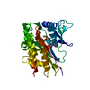
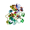
Database: PDB / ID: 3m7m
Title
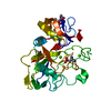
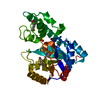
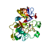
Crystal structure of monomeric hsp33
Components
33 kDa chaperonin
Keywords
CHAPERONE / alpha/beta structure / Disulfide bond / Redox-active center / Stress response
Function / homology
Function and homology information
: / protein folding chaperone / : / response to heat / protein refolding / response to oxidative stress / zinc ion binding / identical protein binding / cytoplasm / cytosol Similarity search - Function
helix hairpin bin / Heat shock protein Hsp33, helix hairpin bin domain superfamily / Hsp33 domain / Hsp33 domain / Heat shock protein Hsp33 / Heat shock protein Hsp33, N-terminal / Heat shock protein Hsp33, C-terminal / Hsp33 protein / 3-Layer(bab) Sandwich / Helix Hairpins ...helix hairpin bin / Heat shock protein Hsp33, helix hairpin bin domain superfamily / Hsp33 domain / Hsp33 domain / Heat shock protein Hsp33 / Heat shock protein Hsp33, N-terminal / Heat shock protein Hsp33, C-terminal / Hsp33 protein / 3-Layer(bab) Sandwich / Helix Hairpins / Orthogonal Bundle / Mainly Alpha / Alpha Beta Similarity search - Domain/homology
In the structure databanks used in Yorodumi, some data are registered as the other names, "COVID-19 virus" and "2019-nCoV". Here are the details of the virus and the list of structure data.
Jan 31, 2019. EMDB accession codes are about to change! (news from PDBe EMDB page)
EMDB accession codes are about to change! (news from PDBe EMDB page)
The allocation of 4 digits for EMDB accession codes will soon come to an end. Whilst these codes will remain in use, new EMDB accession codes will include an additional digit and will expand incrementally as the available range of codes is exhausted. The current 4-digit format prefixed with “EMD-” (i.e. EMD-XXXX) will advance to a 5-digit format (i.e. EMD-XXXXX), and so on. It is currently estimated that the 4-digit codes will be depleted around Spring 2019, at which point the 5-digit format will come into force.
The EM Navigator/Yorodumi systems omit the EMD- prefix.
Related info.:Q: What is EMD? / ID/Accession-code notation in Yorodumi/EM Navigator
Yorodumi is a browser for structure data from EMDB, PDB, SASBDB, etc.
This page is also the successor to EM Navigator detail page, and also detail information page/front-end page for Omokage search.
The word "yorodu" (or yorozu) is an old Japanese word meaning "ten thousand". "mi" (miru) is to see.
Related info.:EMDB / PDB / SASBDB / Comparison of 3 databanks / Yorodumi Search / Aug 31, 2016. New EM Navigator & Yorodumi / Yorodumi Papers / Jmol/JSmol / Function and homology information / Changes in new EM Navigator and Yorodumi
 Movie
Movie Controller


 Open data
 Basic information
 Components
 Keywords
 Function and homology information
 X-RAY DIFFRACTION /
  Authors
 Citation
 Structure visualization
 Downloads & links
 Other downloads
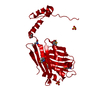


 PDBj
PDBj
 Assembly
 Sample preparation
 / Beamline: 6B / Wavelength: 1 Å
 Processing