 Movie
Movie Controller


+ Open data

- Basic information
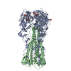
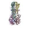
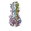
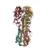
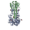
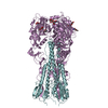
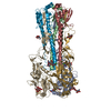
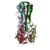
| Entry | Database: PDB / ID: 3m5g | |||||||||
|---|---|---|---|---|---|---|---|---|---|---|
| Title | Crystal structure of a H7 influenza virus hemagglutinin | |||||||||
 Components Components | (Hemagglutinin) x 2 | |||||||||
 Keywords Keywords | VIRAL PROTEIN / influenza virus / hemagglutinin / Envelope protein / Fusion protein / Host cell membrane / Host membrane / Membrane / Transmembrane / Virion | |||||||||
| Function / homology |  Function and homology information Function and homology informationviral budding from plasma membrane / clathrin-dependent endocytosis of virus by host cell / host cell surface receptor binding / fusion of virus membrane with host plasma membrane / fusion of virus membrane with host endosome membrane / viral envelope / virion attachment to host cell / host cell plasma membrane / virion membrane / membrane Similarity search - Function | |||||||||
| Biological species |   Influenza A virus Influenza A virus | |||||||||
| Method |  X-RAY DIFFRACTION / SYNCHROTRON / MOLECULAR REPLACEMENT / Resolution: 2.6 Å X-RAY DIFFRACTION / SYNCHROTRON / MOLECULAR REPLACEMENT / Resolution: 2.6 Å | |||||||||
 Authors Authors | Yang, H. / Chen, L.M. / Carney, P.J. / Donis, R.O. / Stevens, J. | |||||||||
 Citation Citation | Journal: Plos Pathog. / Year: 2010 Title: Structures of receptor complexes of a North American H7N2 influenza hemagglutinin with a loop deletion in the receptor binding site. Authors: Yang, H. / Chen, L.M. / Carney, P.J. / Donis, R.O. / Stevens, J. | |||||||||
| History |
|
- Structure visualization
| Structure viewer | Molecule: MolmilJmol/JSmol |
|---|
- Downloads & links
-Download
| PDBx/mmCIF format | 3m5g.cif.gz | 296.1 KB | Display | PDBx/mmCIF format |
|---|---|---|---|---|
| PDB format | pdb3m5g.ent.gz | 237.9 KB | Display | PDB format |
| PDBx/mmJSON format | 3m5g.json.gz | Tree view | PDBx/mmJSON format | |
| Others |  Other downloads Other downloads |
-Validation report
| Arichive directory | https://data.pdbj.org/pub/pdb/validation_reports/m5/3m5gftp://data.pdbj.org/pub/pdb/validation_reports/m5/3m5g | HTTPS FTP |
|---|
-Related structure data
| Related structure data | 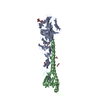 3m5hC 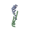 3m5iC  3m5jC  1ti8S C: citing same article ( S: Starting model for refinement |
|---|---|
| Similar structure data |


-Links
 PDBj
PDBj


- Assembly
| Deposited unit | 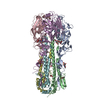
| ||||||||
|---|---|---|---|---|---|---|---|---|---|
| 1 | 
| ||||||||
| Unit cell |
|
-Components
-Protein , 2 types, 6 molecules ACEBDF
| #1: Protein | Mass: 34880.359 Da / Num. of mol.: 3 / Fragment: Hemagglutinin HA1 Source method: isolated from a genetically manipulated source Source: (gene. exp.) Influenza A virus / Production host:  Trichoplusia ni (cabbage looper) / References: UniProt: B7NY59 Trichoplusia ni (cabbage looper) / References: UniProt: B7NY59#2: Protein | Mass: 20895.980 Da / Num. of mol.: 3 / Fragment: Hemagglutinin HA2 Source method: isolated from a genetically manipulated source Source: (gene. exp.) Influenza A virus / Gene: HA / Production host: Trichoplusia ni (cabbage looper) / References: UniProt: B7NYS1 |
|---|
-Sugars , 2 types, 6 molecules 
| #3: Polysaccharide | Source method: isolated from a genetically manipulated source #4: Sugar |  Type: D-saccharide, beta linking / Mass: 221.208 Da / Num. of mol.: 3 Type: D-saccharide, beta linking / Mass: 221.208 Da / Num. of mol.: 3Source method: isolated from a genetically manipulated source Formula: C8H15NO6 |
|---|
-Non-polymers , 2 types, 296 molecules 

| #5: Chemical |  Mass: 106.120 Da / Num. of mol.: 2 / Source method: obtained synthetically / Formula: C4H10O3 Mass: 106.120 Da / Num. of mol.: 2 / Source method: obtained synthetically / Formula: C4H10O3#6: Water | ChemComp-HOH / | Mass: 18.015 Da / Num. of mol.: 294 / Source method: isolated from a natural source / Formula: H2O |
|---|
-Details
| Has protein modification | Y |
|---|---|
| Sequence details | THE CONFLICTS IN THE SEQUENCE IS BECAUSE THE AUTHORS USED THE GENEBANK SEQUENCE ACC55270.1 WHICH IS ...THE CONFLICTS IN THE SEQUENCE IS BECAUSE THE AUTHORS USED THE GENEBANK SEQUENCE ACC55270.1 WHICH IS A SLIGHTLY DIFFERENT STRAIN |
-Experimental details
-Experiment
| Experiment | Method: X-RAY DIFFRACTION / Number of used crystals: 1 |
|---|
- Sample preparation
| Crystal | Density Matthews: 2.89 Å3/Da / Density % sol: 57.51 % |
|---|---|
| Crystal grow | Temperature: 298 K / Method: microbatch under oil / pH: 7.2 Details: 20% PEG 3350, 0.2 M magnesium chloride at pH 7.2, Microbatch under oil, temperature 298K |
-Data collection
| Diffraction | Mean temperature: 100 K |
|---|---|
| Diffraction source | Source: SYNCHROTRON / Site: APS  / Beamline: 22-ID / Wavelength: 1 Å / Beamline: 22-ID / Wavelength: 1 Å |
| Detector | Type: MAR scanner 300 mm plate / Detector: IMAGE PLATE / Date: Aug 12, 2008 |
| Radiation | Protocol: SINGLE WAVELENGTH / Monochromatic (M) / Laue (L): M / Scattering type: x-ray |
| Radiation wavelength | Wavelength: 1 Å / Relative weight: 1 |
| Reflection | Resolution: 2.6→50 Å / Num. all: 62462 / Num. obs: 61857 / % possible obs: 99.2 % / Redundancy: 7.2 % / Rsym value: 0.106 / Net I/σ(I): 39.6 |
| Reflection shell | Resolution: 2.6→2.69 Å / Redundancy: 6.2 % / Mean I/σ(I) obs: 2 / Rsym value: 0.413 / % possible all: 99 |
- Processing
| Software |
| |||||||||||||||||||||||||||||||||||||||||||||||||||||||||||||||||||||||||||||||||||||||||||||||||||||||||||||||||||||||||||||||||||||||||||||||||||||||||||||||||||||||||||||||
|---|---|---|---|---|---|---|---|---|---|---|---|---|---|---|---|---|---|---|---|---|---|---|---|---|---|---|---|---|---|---|---|---|---|---|---|---|---|---|---|---|---|---|---|---|---|---|---|---|---|---|---|---|---|---|---|---|---|---|---|---|---|---|---|---|---|---|---|---|---|---|---|---|---|---|---|---|---|---|---|---|---|---|---|---|---|---|---|---|---|---|---|---|---|---|---|---|---|---|---|---|---|---|---|---|---|---|---|---|---|---|---|---|---|---|---|---|---|---|---|---|---|---|---|---|---|---|---|---|---|---|---|---|---|---|---|---|---|---|---|---|---|---|---|---|---|---|---|---|---|---|---|---|---|---|---|---|---|---|---|---|---|---|---|---|---|---|---|---|---|---|---|---|---|---|---|---|
| Refinement | Method to determine structure: MOLECULAR REPLACEMENT Starting model: PDB Entry 1TI8 Resolution: 2.6→50 Å / Cor.coef. Fo:Fc: 0.917 / Cor.coef. Fo:Fc free: 0.882 / SU B: 20.843 / SU ML: 0.215 / Cross valid method: THROUGHOUT / ESU R: 0.621 / ESU R Free: 0.308 / Stereochemistry target values: MAXIMUM LIKELIHOOD / Details: HYDROGENS HAVE BEEN ADDED IN THE RIDING POSITIONS
| |||||||||||||||||||||||||||||||||||||||||||||||||||||||||||||||||||||||||||||||||||||||||||||||||||||||||||||||||||||||||||||||||||||||||||||||||||||||||||||||||||||||||||||||
| Solvent computation | Ion probe radii: 0.8 Å / Shrinkage radii: 0.8 Å / VDW probe radii: 1.2 Å / Solvent model: MASK | |||||||||||||||||||||||||||||||||||||||||||||||||||||||||||||||||||||||||||||||||||||||||||||||||||||||||||||||||||||||||||||||||||||||||||||||||||||||||||||||||||||||||||||||
| Displacement parameters | Biso mean: 16.926 Å2
| |||||||||||||||||||||||||||||||||||||||||||||||||||||||||||||||||||||||||||||||||||||||||||||||||||||||||||||||||||||||||||||||||||||||||||||||||||||||||||||||||||||||||||||||
| Refinement step | Cycle: LAST / Resolution: 2.6→50 Å
| |||||||||||||||||||||||||||||||||||||||||||||||||||||||||||||||||||||||||||||||||||||||||||||||||||||||||||||||||||||||||||||||||||||||||||||||||||||||||||||||||||||||||||||||
| Refine LS restraints |
| |||||||||||||||||||||||||||||||||||||||||||||||||||||||||||||||||||||||||||||||||||||||||||||||||||||||||||||||||||||||||||||||||||||||||||||||||||||||||||||||||||||||||||||||
| LS refinement shell | Resolution: 2.6→2.667 Å / Total num. of bins used: 20
| |||||||||||||||||||||||||||||||||||||||||||||||||||||||||||||||||||||||||||||||||||||||||||||||||||||||||||||||||||||||||||||||||||||||||||||||||||||||||||||||||||||||||||||||
| Refinement TLS params. | Method: refined / Refine-ID: X-RAY DIFFRACTION
| |||||||||||||||||||||||||||||||||||||||||||||||||||||||||||||||||||||||||||||||||||||||||||||||||||||||||||||||||||||||||||||||||||||||||||||||||||||||||||||||||||||||||||||||
| Refinement TLS group |
|
