 Movie
Movie Controller


+ Open data

- Basic information
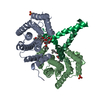
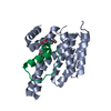
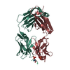
| Entry | Database: PDB / ID: 3m51 | ||||||
|---|---|---|---|---|---|---|---|
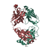
| Title | Structure of the 14-3-3/PMA2 complex stabilized by Pyrrolidone1 | ||||||
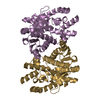
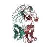
 Components Components |
| ||||||
 Keywords Keywords | PROTEIN BINDING / all helical / protein-protein complex | ||||||
| Function / homology |  Function and homology information Function and homology informationP-type H+-exporting transporter / proton export across plasma membrane / P-type proton-exporting transporter activity / intracellular protein localization / signal transduction / ATP hydrolysis activity / ATP binding / metal ion binding / plasma membrane / cytoplasm Similarity search - Function | ||||||
| Biological species |  | ||||||
| Method |  X-RAY DIFFRACTION / SYNCHROTRON / MOLECULAR REPLACEMENT / molecular replacement / Resolution: 3.25 Å X-RAY DIFFRACTION / SYNCHROTRON / MOLECULAR REPLACEMENT / molecular replacement / Resolution: 3.25 Å | ||||||
 Authors Authors | Ottmann, C. / Rose, R. / Waldmann, H. | ||||||
 Citation Citation | Journal: Angew.Chem.Int.Ed.Engl. / Year: 2010 Title: Identification and structure of small-molecule stabilizers of 14-3-3 protein-protein interactions Authors: Rose, R. / Erdmann, S. / Bovens, S. / Wolf, A. / Rose, M. / Hennig, S. / Waldmann, H. / Ottmann, C. | ||||||
| History |
|
- Structure visualization
| Structure viewer | Molecule: MolmilJmol/JSmol |
|---|
- Downloads & links
-Download
| PDBx/mmCIF format | 3m51.cif.gz | 63.9 KB | Display | PDBx/mmCIF format |
|---|---|---|---|---|
| PDB format | pdb3m51.ent.gz | 46.8 KB | Display | PDB format |
| PDBx/mmJSON format | 3m51.json.gz | Tree view | PDBx/mmJSON format | |
| Others |  Other downloads Other downloads |
-Validation report
| Arichive directory | https://data.pdbj.org/pub/pdb/validation_reports/m5/3m51ftp://data.pdbj.org/pub/pdb/validation_reports/m5/3m51 | HTTPS FTP |
|---|
-Related structure data
| Related structure data |  3m50C 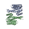 1yz5S C: citing same article ( S: Starting model for refinement |
|---|---|
| Similar structure data |
-Links
 PDBj
PDBj


- Assembly
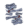
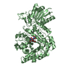
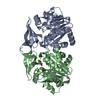
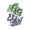
| Deposited unit | 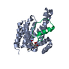
| ||||||||
|---|---|---|---|---|---|---|---|---|---|
| 1 | 
| ||||||||
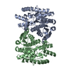
| Unit cell |
|
-Components
| #1: Protein | Mass: 27169.635 Da / Num. of mol.: 1 / Fragment: residues 1-240 Source method: isolated from a genetically manipulated source Source: (gene. exp.)  |
|---|---|
| #2: Protein/peptide | Mass: 3597.148 Da / Num. of mol.: 1 / Fragment: C-terminal fragment / Mutation: S938A,T955D,V956I Source method: isolated from a genetically manipulated source Source: (gene. exp.) Plasmid: PTYB12 / Production host: |
| #3: Chemical | ChemComp-YR1 / 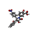  Mass: 460.392 Da / Num. of mol.: 1 / Source method: obtained synthetically / Formula: C24H16N2O8 Mass: 460.392 Da / Num. of mol.: 1 / Source method: obtained synthetically / Formula: C24H16N2O8 |
| #4: Water | ChemComp-HOH /  Mass: 18.015 Da / Num. of mol.: 7 / Source method: isolated from a natural source / Formula: H2O Mass: 18.015 Da / Num. of mol.: 7 / Source method: isolated from a natural source / Formula: H2O |
-Experimental details
-Experiment
| Experiment | Method: X-RAY DIFFRACTION / Number of used crystals: 1 |
|---|
- Sample preparation
| Crystal | Density Matthews: 4.13 Å3/Da / Density % sol: 70.24 % |
|---|---|
| Crystal grow | Temperature: 277.15 K / Method: vapor diffusion, hanging drop / pH: 9 Details: 0.1M CHES, 1.0M Na-Citrat, 30%(w/v) sucrose, pH 9.0, VAPOR DIFFUSION, HANGING DROP, temperature 277.15K |
-Data collection
| Diffraction | Mean temperature: 100 K | ||||||||||||||||||||||||||||||||||||||||||||||||||||||||
|---|---|---|---|---|---|---|---|---|---|---|---|---|---|---|---|---|---|---|---|---|---|---|---|---|---|---|---|---|---|---|---|---|---|---|---|---|---|---|---|---|---|---|---|---|---|---|---|---|---|---|---|---|---|---|---|---|---|
| Diffraction source | Source: SYNCHROTRON / Site: SLS  / Beamline: X10SA / Wavelength: 1.00749 Å / Beamline: X10SA / Wavelength: 1.00749 Å | ||||||||||||||||||||||||||||||||||||||||||||||||||||||||
| Detector | Type: MARMOSAIC 225 mm CCD / Detector: CCD / Date: Apr 4, 2009 | ||||||||||||||||||||||||||||||||||||||||||||||||||||||||
| Radiation | Protocol: SINGLE WAVELENGTH / Monochromatic (M) / Laue (L): M / Scattering type: x-ray | ||||||||||||||||||||||||||||||||||||||||||||||||||||||||
| Radiation wavelength | Wavelength: 1.00749 Å / Relative weight: 1 | ||||||||||||||||||||||||||||||||||||||||||||||||||||||||
| Reflection | Resolution: 3.25→48.74 Å / Num. obs: 8499 / % possible obs: 99.9 % / Observed criterion σ(I): -3 / Biso Wilson estimate: 85.969 Å2 / Rmerge(I) obs: 0.061 / Net I/σ(I): 22.39 | ||||||||||||||||||||||||||||||||||||||||||||||||||||||||
| Reflection shell | Diffraction-ID: 1
|
-Phasing
| Phasing | Method: molecular replacement |
|---|
- Processing
| Software |
| |||||||||||||||||||||||||||||||||||||||||||||||||||||||||||||||||
|---|---|---|---|---|---|---|---|---|---|---|---|---|---|---|---|---|---|---|---|---|---|---|---|---|---|---|---|---|---|---|---|---|---|---|---|---|---|---|---|---|---|---|---|---|---|---|---|---|---|---|---|---|---|---|---|---|---|---|---|---|---|---|---|---|---|---|
| Refinement | Method to determine structure: MOLECULAR REPLACEMENT Starting model: 1YZ5 Resolution: 3.25→48.74 Å / Cor.coef. Fo:Fc: 0.899 / Cor.coef. Fo:Fc free: 0.884 / WRfactor Rfree: 0.324 / WRfactor Rwork: 0.295 / Occupancy max: 1 / Occupancy min: 0.5 / FOM work R set: 0.737 / SU Rfree: 0.567 / Cross valid method: THROUGHOUT / σ(F): 0 / ESU R Free: 0.567 / Stereochemistry target values: MAXIMUM LIKELIHOOD Details: HYDROGENS HAVE BEEN ADDED IN THE RIDING POSITIONS U VALUES: REFINED INDIVIDUALLY
| |||||||||||||||||||||||||||||||||||||||||||||||||||||||||||||||||
| Solvent computation | Ion probe radii: 0.8 Å / Shrinkage radii: 0.8 Å / VDW probe radii: 1.4 Å / Solvent model: MASK | |||||||||||||||||||||||||||||||||||||||||||||||||||||||||||||||||
| Displacement parameters | Biso max: 116.37 Å2 / Biso mean: 98.388 Å2 / Biso min: 54.38 Å2
| |||||||||||||||||||||||||||||||||||||||||||||||||||||||||||||||||
| Refinement step | Cycle: LAST / Resolution: 3.25→48.74 Å
| |||||||||||||||||||||||||||||||||||||||||||||||||||||||||||||||||
| Refine LS restraints |
| |||||||||||||||||||||||||||||||||||||||||||||||||||||||||||||||||
| LS refinement shell | Resolution: 3.25→3.334 Å / Total num. of bins used: 20
|
