 Movie
Movie Controller

[English] 日本語
 Yorodumi
- PDB-3m0r: Crystal Structure of the R21D mutant of alpha-spectrin SH3 domain... -
+ Open data

- Basic information
| Entry | Database: PDB / ID: 3m0r | ||||||
|---|---|---|---|---|---|---|---|
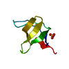
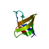
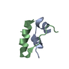
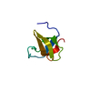
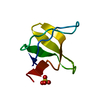
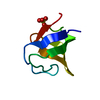
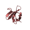
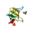
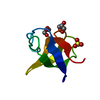
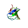
| Title | Crystal Structure of the R21D mutant of alpha-spectrin SH3 domain. Crystal obtained in ammonium sulphate at pH 6. | ||||||
 Components Components | Spectrin alpha chain, brain | ||||||
 Keywords Keywords | SIGNALING PROTEIN / SH3-like barrel / Actin capping / Actin-binding / Calmodulin-binding / Cytoskeleton / Phosphoprotein / SH3 domain | ||||||
| Function / homology |  Function and homology information Function and homology informationcostamere / actin filament capping / cortical actin cytoskeleton / cell projection / cell junction / actin filament binding / actin cytoskeleton organization / calmodulin binding / calcium ion binding / plasma membrane Similarity search - Function | ||||||
| Biological species |  | ||||||
| Method |  X-RAY DIFFRACTION / SYNCHROTRON / MOLECULAR REPLACEMENT / molecular replacement / Resolution: 1.1 Å X-RAY DIFFRACTION / SYNCHROTRON / MOLECULAR REPLACEMENT / molecular replacement / Resolution: 1.1 Å | ||||||
 Authors Authors | Gavira, J.A. / Camara-Artigas, A. | ||||||
 Citation Citation | Journal: Acta Crystallogr.,Sect.D / Year: 2011 Title: Understanding the polymorphic behaviour of a mutant of the alpha-spectrin SH3 domain by means of two 1.1 A structures Authors: Camara-Artigas, A. / Gavira, J.A. / Casares, S. / Garcia-Ruiz, J.M. / Conejero-Lara, F. / Allen, J.P. / Martinez, J. | ||||||
| History |
|
- Structure visualization
| Structure viewer | Molecule: MolmilJmol/JSmol |
|---|
- Downloads & links
-Download
| PDBx/mmCIF format | 3m0r.cif.gz | 42 KB | Display | PDBx/mmCIF format |
|---|---|---|---|---|
| PDB format | pdb3m0r.ent.gz | 28.8 KB | Display | PDB format |
| PDBx/mmJSON format | 3m0r.json.gz | Tree view | PDBx/mmJSON format | |
| Others |  Other downloads Other downloads |
-Validation report
| Arichive directory | https://data.pdbj.org/pub/pdb/validation_reports/m0/3m0rftp://data.pdbj.org/pub/pdb/validation_reports/m0/3m0r | HTTPS FTP |
|---|
-Related structure data
| Related structure data |  3m0pC 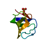 3m0qC  3m0sC  3m0tC 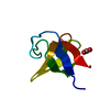 3m0uC 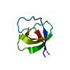 1shgS S: Starting model for refinement C: citing same article ( |
|---|---|
| Similar structure data |


-Links
 PDBj
PDBj- Assembly
| Deposited unit | 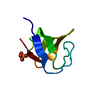
| ||||||||
|---|---|---|---|---|---|---|---|---|---|
| 1 |
| ||||||||
| Unit cell |
|
-Components
| #1: Protein | Mass: 7187.138 Da / Num. of mol.: 1 / Fragment: SH3 domain (UNP residues 965 to 1025) / Mutation: R21D Source method: isolated from a genetically manipulated source Source: (gene. exp.)  |
|---|---|
| #2: Chemical | ChemComp-SO4 /   Mass: 96.063 Da / Num. of mol.: 1 / Source method: obtained synthetically / Formula: SO4 Mass: 96.063 Da / Num. of mol.: 1 / Source method: obtained synthetically / Formula: SO4 |
| #3: Water | ChemComp-HOH /  Mass: 18.015 Da / Num. of mol.: 109 / Source method: isolated from a natural source / Formula: H2O Mass: 18.015 Da / Num. of mol.: 109 / Source method: isolated from a natural source / Formula: H2O |
-Experimental details
-Experiment
| Experiment | Method: X-RAY DIFFRACTION / Number of used crystals: 1 |
|---|
- Sample preparation
| Crystal | Density Matthews: 2.48 Å3/Da / Density % sol: 50.32 % |
|---|---|
| Crystal grow | Temperature: 298 K / Method: vapor diffusion, hanging drop / pH: 6 Details: 0.8 M ammonium sulphate, 0.1 M Bis-Tris pH 6, VAPOR DIFFUSION, HANGING DROP, temperature 298K |
-Data collection
| Diffraction | Mean temperature: 100 K |
|---|---|
| Diffraction source | Source: SYNCHROTRON / Site: ESRF  / Beamline: BM16 / Wavelength: 1.033 Å / Beamline: BM16 / Wavelength: 1.033 Å |
| Detector | Type: CCD ADSC_Q210 / Detector: CCD / Date: Apr 4, 2009 |
| Radiation | Protocol: SINGLE WAVELENGTH / Monochromatic (M) / Laue (L): M / Scattering type: x-ray |
| Radiation wavelength | Wavelength: 1.033 Å / Relative weight: 1 |
| Reflection | Resolution: 1.1→31.799 Å / Num. all: 29563 / Num. obs: 28144 / % possible obs: 95.2 % / Observed criterion σ(F): 0 / Observed criterion σ(I): 0 / Redundancy: 6.7 % / Biso Wilson estimate: 36.394 Å2 / Rmerge(I) obs: 0.091 / Rsym value: 0.091 / Net I/σ(I): 16.8 |
| Reflection shell | Resolution: 1.1→1.14 Å / Redundancy: 6.7 % / Rmerge(I) obs: 0.302 / Mean I/σ(I) obs: 6.4 / Num. unique all: 2867 / Rsym value: 0.302 / % possible all: 98.8 |
-Phasing
| Phasing | Method: molecular replacement | |||||||||
|---|---|---|---|---|---|---|---|---|---|---|
| Phasing MR |
|
- Processing
| Software |
| ||||||||||||||||||||||||||||||||||||||||||||||||||||||||||||||||||||||||||||||||
|---|---|---|---|---|---|---|---|---|---|---|---|---|---|---|---|---|---|---|---|---|---|---|---|---|---|---|---|---|---|---|---|---|---|---|---|---|---|---|---|---|---|---|---|---|---|---|---|---|---|---|---|---|---|---|---|---|---|---|---|---|---|---|---|---|---|---|---|---|---|---|---|---|---|---|---|---|---|---|---|---|---|
| Refinement | Method to determine structure: MOLECULAR REPLACEMENT Starting model: PDB entry 1SHG Resolution: 1.1→18.05 Å / Cor.coef. Fo:Fc: 0.956 / Cor.coef. Fo:Fc free: 0.944 / WRfactor Rfree: 0.198 / WRfactor Rwork: 0.176 / Occupancy max: 1 / Occupancy min: 0.5 / FOM work R set: 0.9 / SU B: 0.76 / SU ML: 0.017 / SU R Cruickshank DPI: 0.032 / SU Rfree: 0.033 / Cross valid method: THROUGHOUT / σ(F): 0 / σ(I): 0 / ESU R: 0.032 / ESU R Free: 0.033 / Stereochemistry target values: MAXIMUM LIKELIHOOD Details: HYDROGENS HAVE BEEN ADDED IN THE RIDING POSITIONS U VALUES : REFINED INDIVIDUALLY
| ||||||||||||||||||||||||||||||||||||||||||||||||||||||||||||||||||||||||||||||||
| Solvent computation | Ion probe radii: 0.8 Å / Shrinkage radii: 0.8 Å / VDW probe radii: 1.4 Å / Solvent model: MASK | ||||||||||||||||||||||||||||||||||||||||||||||||||||||||||||||||||||||||||||||||
| Displacement parameters | Biso max: 47.49 Å2 / Biso mean: 14.168 Å2 / Biso min: 6.17 Å2
| ||||||||||||||||||||||||||||||||||||||||||||||||||||||||||||||||||||||||||||||||
| Refinement step | Cycle: LAST / Resolution: 1.1→18.05 Å
| ||||||||||||||||||||||||||||||||||||||||||||||||||||||||||||||||||||||||||||||||
| Refine LS restraints |
| ||||||||||||||||||||||||||||||||||||||||||||||||||||||||||||||||||||||||||||||||
| LS refinement shell | Resolution: 1.102→1.13 Å / Total num. of bins used: 20
|
