- PDB-3lyp: Structure of stringent starvation protein A homolog from Pseudomo... -
+
Open data
ID or keywords:
Loading...
-
Basic information
Entry
Database: PDB / ID: 3lyp
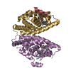
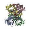
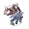
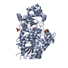
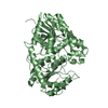
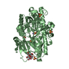
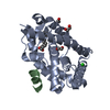
Title
Structure of stringent starvation protein A homolog from Pseudomonas fluorescens
Components
Stringent starvation protein A
Keywords
TRANSCRIPTION / structural genomics / GST-superfamily / sspA / Stringent starvation protein A homolog / PSI-2 / Protein Structure Initiative / New York SGX Research Center for Structural Genomics / NYSGXRC
Resolution: 1.6→50 Å / Cor.coef. Fo:Fc: 0.954 / Cor.coef. Fo:Fc free: 0.941 / WRfactor Rfree: 0.229 / WRfactor Rwork: 0.195 / Occupancy max: 1 / Occupancy min: 0.3 / FOM work R set: 0.881 / SU B: 3.088 / SU ML: 0.051 / SU R Cruickshank DPI: 0.093 / SU Rfree: 0.092 / TLS residual ADP flag: LIKELY RESIDUAL / Cross valid method: THROUGHOUT / σ(F): 0 / ESU R: 0.093 / ESU R Free: 0.092 / Stereochemistry target values: MAXIMUM LIKELIHOOD Details: HYDROGENS HAVE BEEN ADDED IN THE RIDING POSITIONS, U VALUES RESIDUAL ONLY, UNCHARACTERIZED ELECTRON DENSITY OBSERVED BETWEEN SIDE-CHAINS OF THE RESIDUES ARG-103 AND TRP-108 (CHAIN A and B). ...Details: HYDROGENS HAVE BEEN ADDED IN THE RIDING POSITIONS, U VALUES RESIDUAL ONLY, UNCHARACTERIZED ELECTRON DENSITY OBSERVED BETWEEN SIDE-CHAINS OF THE RESIDUES ARG-103 AND TRP-108 (CHAIN A and B). DENSITY SIMILAR TO PEPTIDE CONTAINING GLY-ASN/ASP OBSERVED NEAR RESIDUES 28-29 OF CHAIN B IS NOT MODELLED.
Rfactor
Num. reflection
% reflection
Selection details
Rfree
0.212
2741
5.1 %
RANDOM
Rwork
0.183
-
-
-
obs
0.185
54182
95.44 %
-
Solvent computation
Ion probe radii: 0.8 Å / Shrinkage radii: 0.8 Å / VDW probe radii: 1.4 Å / Solvent model: MASK
In the structure databanks used in Yorodumi, some data are registered as the other names, "COVID-19 virus" and "2019-nCoV". Here are the details of the virus and the list of structure data.
Jan 31, 2019. EMDB accession codes are about to change! (news from PDBe EMDB page)
EMDB accession codes are about to change! (news from PDBe EMDB page)
The allocation of 4 digits for EMDB accession codes will soon come to an end. Whilst these codes will remain in use, new EMDB accession codes will include an additional digit and will expand incrementally as the available range of codes is exhausted. The current 4-digit format prefixed with “EMD-” (i.e. EMD-XXXX) will advance to a 5-digit format (i.e. EMD-XXXXX), and so on. It is currently estimated that the 4-digit codes will be depleted around Spring 2019, at which point the 5-digit format will come into force.
The EM Navigator/Yorodumi systems omit the EMD- prefix.
Related info.:Q: What is EMD? / ID/Accession-code notation in Yorodumi/EM Navigator
Yorodumi is a browser for structure data from EMDB, PDB, SASBDB, etc.
This page is also the successor to EM Navigator detail page, and also detail information page/front-end page for Omokage search.
The word "yorodu" (or yorozu) is an old Japanese word meaning "ten thousand". "mi" (miru) is to see.
Related info.:EMDB / PDB / SASBDB / Comparison of 3 databanks / Yorodumi Search / Aug 31, 2016. New EM Navigator & Yorodumi / Yorodumi Papers / Jmol/JSmol / Function and homology information / Changes in new EM Navigator and Yorodumi
 Movie
Movie Controller

 Yorodumi
 Open data
 Basic information
 Components
 Keywords
 Function and homology information
 Pseudomonas fluorescens (bacteria)
 X-RAY DIFFRACTION /
  Authors
 Citation
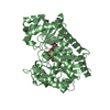
 Structure visualization
 Downloads & links
 Other downloads
 PDBj
PDBj

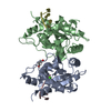
 Assembly
 Mass: 18.015 Da / Num. of mol.: 217 / Source method: isolated from a natural source / Formula: H2O
 Sample preparation
 / Beamline: X29A / Wavelength: 0.9793 Å
 Processing