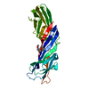
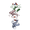
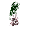
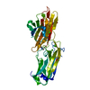
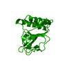
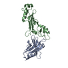
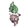
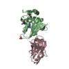
Mass: 29164.166 Da / Num. of mol.: 1 / Fragment: L27 domain, L27 1/2 domain Source method: isolated from a genetically manipulated source Details: Structural Basis for Assembling a Tripartite Complex Dlg1-MPP7-Mals3 Source: (gene. exp.) Homo sapiens (human) / Gene: L27 domains of Dlg1, MPP7 and Mals3 Plasmid details: in-house-modified version of the pET32a vector (Novagen), in which the S-tag an d the thrombin recognition site were replaced by a sequence encoding a TEV prote ase cleavage site ...Plasmid details: in-house-modified version of the pET32a vector (Novagen), in which the S-tag an d the thrombin recognition site were replaced by a sequence encoding a TEV prote ase cleavage site (Glu-Asn-Leu-Tyr-Phe-Gln-Ser) Plasmid: pET32a vector (Novagen) / Production host: Escherichia coli (E. coli) / Strain (production host): BL21(DE3) References: UniProt: Q12959, UniProt: Q5T2T1, UniProt: Q9NUP9
Mass: 18.015 Da / Num. of mol.: 41 / Source method: isolated from a natural source / Formula: H2O
Compound details
THE L27 DOMAINS OF HUMAN DLG1 (TERMED AS L27HDLG1), HUMAN MPP7 (TERMED AS L27N AND L27C) AND HUMAN ...THE L27 DOMAINS OF HUMAN DLG1 (TERMED AS L27HDLG1), HUMAN MPP7 (TERMED AS L27N AND L27C) AND HUMAN MALS3 (TERMED AS L27HMALS3) WERE COVALENTLY LINKED TOGETHER BY PRECISSION PROTEASE CLEAVAGE SITE AS ONE SINGLE POLYPEPTIDE.
Sequence details
THE INTERNAL SEQUENCE LEVLFQGP ARE LINKER.
-
Experimental details
-
Experiment
Experiment
Method: X-RAY DIFFRACTION / Number of used crystals: 2
-
Sample preparation
Crystal
Density Matthews: 3.42 Å3/Da / Density % sol: 64.04 %
Crystal grow
Temperature: 293 K / Method: vapor diffusion, sitting drop / pH: 7 Details: 10% PEG 8000, 0.2M MgCl2, 0.1M Guanidine HCl, 0.1M Tris-HCl, pH7.0, VAPOR DIFFUSION, SITTING DROP, temperature 293K
In the structure databanks used in Yorodumi, some data are registered as the other names, "COVID-19 virus" and "2019-nCoV". Here are the details of the virus and the list of structure data.
Jan 31, 2019. EMDB accession codes are about to change! (news from PDBe EMDB page)
EMDB accession codes are about to change! (news from PDBe EMDB page)
The allocation of 4 digits for EMDB accession codes will soon come to an end. Whilst these codes will remain in use, new EMDB accession codes will include an additional digit and will expand incrementally as the available range of codes is exhausted. The current 4-digit format prefixed with “EMD-” (i.e. EMD-XXXX) will advance to a 5-digit format (i.e. EMD-XXXXX), and so on. It is currently estimated that the 4-digit codes will be depleted around Spring 2019, at which point the 5-digit format will come into force.
The EM Navigator/Yorodumi systems omit the EMD- prefix.
Related info.:Q: What is EMD? / ID/Accession-code notation in Yorodumi/EM Navigator
Yorodumi is a browser for structure data from EMDB, PDB, SASBDB, etc.
This page is also the successor to EM Navigator detail page, and also detail information page/front-end page for Omokage search.
The word "yorodu" (or yorozu) is an old Japanese word meaning "ten thousand". "mi" (miru) is to see.
Related info.:EMDB / PDB / SASBDB / Comparison of 3 databanks / Yorodumi Search / Aug 31, 2016. New EM Navigator & Yorodumi / Yorodumi Papers / Jmol/JSmol / Function and homology information / Changes in new EM Navigator and Yorodumi
 Movie
Movie Controller

 Yorodumi
 Open data
 Basic information
 Components
 Keywords
 Function and homology information
 Homo sapiens (human)
 X-RAY DIFFRACTION /
  Authors
 Citation
 Structure visualization
 Downloads & links
 Other downloads
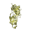

 PDBj
PDBj


 Assembly
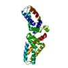

 Mass: 18.015 Da / Num. of mol.: 41 / Source method: isolated from a natural source / Formula: H2O
 Sample preparation

 Processing