 Movie
Movie Controller

[English] 日本語
 Yorodumi
- PDB-2vzd: Crystal structure of the C-terminal calponin homology domain of a... -
+ Open data

- Basic information
| Entry | Database: PDB / ID: 2vzd | ||||||
|---|---|---|---|---|---|---|---|
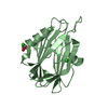
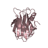
| Title | Crystal structure of the C-terminal calponin homology domain of alpha parvin in complex with paxillin LD1 motif | ||||||
 Components Components |
| ||||||
 Keywords Keywords | CELL ADHESION / CALPONIN HOMOLOGY DOMAIN / CELL MEMBRANE / METAL-BINDING / CYTOSKELETON / CELL JUNCTION / ACTIN-BINDING / PHOSPHOPROTEIN / MEMBRANE / LD1 MOTIF / LIM DOMAIN | ||||||
| Function / homology |  Function and homology information Function and homology informationactin-mediated cell contraction / smooth muscle cell chemotaxis / establishment or maintenance of cell polarity regulating cell shape / Regulation of cytoskeletal remodeling and cell spreading by IPP complex components / Localization of the PINCH-ILK-PARVIN complex to focal adhesions / Regulation of MITF-M-dependent genes involved in extracellular matrix, focal adhesion and epithelial-to-mesenchymal transition / neuropilin binding / vinculin binding / Cell-extracellular matrix interactions / signal complex assembly ...actin-mediated cell contraction / smooth muscle cell chemotaxis / establishment or maintenance of cell polarity regulating cell shape / Regulation of cytoskeletal remodeling and cell spreading by IPP complex components / Localization of the PINCH-ILK-PARVIN complex to focal adhesions / Regulation of MITF-M-dependent genes involved in extracellular matrix, focal adhesion and epithelial-to-mesenchymal transition / neuropilin binding / vinculin binding / Cell-extracellular matrix interactions / signal complex assembly / outflow tract septum morphogenesis / sprouting angiogenesis / microtubule associated complex / heterotypic cell-cell adhesion / growth hormone receptor signaling pathway / protein kinase inhibitor activity / establishment or maintenance of cell polarity / cilium assembly / Smooth Muscle Contraction / GAB1 signalosome / endothelial cell migration / PTK6 Regulates RHO GTPases, RAS GTPase and MAP kinases / positive regulation of stress fiber assembly / stress fiber / substrate adhesion-dependent cell spreading / transforming growth factor beta receptor signaling pathway / cellular response to reactive oxygen species / beta-catenin binding / VEGFA-VEGFR2 Pathway / Z disc / cell-cell junction / cell migration / lamellipodium / actin cytoskeleton / actin binding / actin cytoskeleton organization / protein phosphatase binding / cell cortex / cell adhesion / protein stabilization / cadherin binding / focal adhesion / signal transduction / metal ion binding / plasma membrane / cytoplasm / cytosol Similarity search - Function | ||||||
| Biological species |  HOMO SAPIENS (human) HOMO SAPIENS (human) | ||||||
| Method |  X-RAY DIFFRACTION / SYNCHROTRON / MOLECULAR REPLACEMENT / Resolution: 2.1 Å X-RAY DIFFRACTION / SYNCHROTRON / MOLECULAR REPLACEMENT / Resolution: 2.1 Å | ||||||
 Authors Authors | Lorenz, S. / Vakonakis, I. / Lowe, E.D. / Campbell, I.D. / Noble, M.E.M. / Hoellerer, M.K. | ||||||
 Citation Citation | Journal: Structure / Year: 2008 Title: Structural Analysis of the Interactions between Paxillin Ld Motifs and Alpha-Parvin Authors: Lorenz, S. / Vakonakis, I. / Lowe, E.D. / Campbell, I.D. / Noble, M.E.M. / Hoellerer, M.K. | ||||||
| History |
|
- Structure visualization
| Structure viewer | Molecule: MolmilJmol/JSmol |
|---|
- Downloads & links
-Download
| PDBx/mmCIF format | 2vzd.cif.gz | 82.8 KB | Display | PDBx/mmCIF format |
|---|---|---|---|---|
| PDB format | pdb2vzd.ent.gz | 64.4 KB | Display | PDB format |
| PDBx/mmJSON format | 2vzd.json.gz | Tree view | PDBx/mmJSON format | |
| Others |  Other downloads Other downloads |
-Validation report
| Arichive directory | https://data.pdbj.org/pub/pdb/validation_reports/vz/2vzdftp://data.pdbj.org/pub/pdb/validation_reports/vz/2vzd | HTTPS FTP |
|---|
-Related structure data
| Related structure data |  2vzcSC 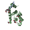 2vzgC 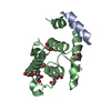 2vziC C: citing same article ( S: Starting model for refinement |
|---|---|
| Similar structure data |


-Links
 PDBj
PDBj


- Assembly
| Deposited unit | 
| |||||||||||||||||||||||||||||||||||||||||||||||||||||||||||
|---|---|---|---|---|---|---|---|---|---|---|---|---|---|---|---|---|---|---|---|---|---|---|---|---|---|---|---|---|---|---|---|---|---|---|---|---|---|---|---|---|---|---|---|---|---|---|---|---|---|---|---|---|---|---|---|---|---|---|---|---|
| 1 | 
| |||||||||||||||||||||||||||||||||||||||||||||||||||||||||||
| 2 | 
| |||||||||||||||||||||||||||||||||||||||||||||||||||||||||||
| Unit cell |
| |||||||||||||||||||||||||||||||||||||||||||||||||||||||||||
| Noncrystallographic symmetry (NCS) | NCS domain:
NCS domain segments: Ens-ID: 1 / Refine code: 4
NCS oper: (Code: given Matrix: (-0.2002, 0.867, -0.4563), Vector: |
-Components
-Protein / Protein/peptide , 2 types, 4 molecules ABCD
| #1: Protein | Mass: 15155.380 Da / Num. of mol.: 2 Fragment: C-TERMINAL CALPONIN HOMOLOGY DOMAIN, RESIDUES 242-372 Source method: isolated from a genetically manipulated source Source: (gene. exp.) HOMO SAPIENS (human) / Plasmid: PGEX-6P1 / Production host:  #2: Protein/peptide | Mass: 2177.388 Da / Num. of mol.: 2 / Fragment: PAXILLIN LD1 MOTIF, RESIDUES 1-20 / Source method: obtained synthetically / Source: (synth.) HOMO SAPIENS (human) / References: UniProt: P49023 |
|---|
-Non-polymers , 5 types, 99 molecules 


| #3: Chemical |  Mass: 150.173 Da / Num. of mol.: 2 / Source method: obtained synthetically / Formula: C6H14O4 Mass: 150.173 Da / Num. of mol.: 2 / Source method: obtained synthetically / Formula: C6H14O4#4: Chemical |  Mass: 194.226 Da / Num. of mol.: 2 / Source method: obtained synthetically / Formula: C8H18O5 / Comment: precipitant*YM Mass: 194.226 Da / Num. of mol.: 2 / Source method: obtained synthetically / Formula: C8H18O5 / Comment: precipitant*YM#5: Chemical |  Mass: 92.094 Da / Num. of mol.: 3 / Source method: obtained synthetically / Formula: C3H8O3 Mass: 92.094 Da / Num. of mol.: 3 / Source method: obtained synthetically / Formula: C3H8O3#6: Chemical | ChemComp-EDO /  Mass: 62.068 Da / Num. of mol.: 7 / Source method: obtained synthetically / Formula: C2H6O2 Mass: 62.068 Da / Num. of mol.: 7 / Source method: obtained synthetically / Formula: C2H6O2#7: Water | ChemComp-HOH / | Mass: 18.015 Da / Num. of mol.: 85 / Source method: isolated from a natural source / Formula: H2O |
|---|
-Experimental details
-Experiment
| Experiment | Method: X-RAY DIFFRACTION / Number of used crystals: 1 |
|---|
- Sample preparation
| Crystal | Density Matthews: 2.6 Å3/Da / Density % sol: 52.36 % / Description: NONE |
|---|---|
| Crystal grow | pH: 7.5 / Details: 20% (W/V) PEG 10000, 0.1M HEPES PH 7.5 |
-Data collection
| Diffraction | Mean temperature: 100 K |
|---|---|
| Diffraction source | Source: SYNCHROTRON / Site: ESRF  / Beamline: ID23-1 / Wavelength: 0.969 / Beamline: ID23-1 / Wavelength: 0.969 |
| Detector | Type: ADSC CCD / Detector: CCD / Date: Nov 5, 2006 / Details: MIRRORS |
| Radiation | Monochromator: SI (111) / Protocol: SINGLE WAVELENGTH / Monochromatic (M) / Laue (L): M / Scattering type: x-ray |
| Radiation wavelength | Wavelength: 0.969 Å / Relative weight: 1 |
| Reflection | Resolution: 2.1→30.03 Å / Num. obs: 20260 / % possible obs: 98.5 % / Observed criterion σ(I): 2 / Redundancy: 2.9 % / Rmerge(I) obs: 0.05 / Net I/σ(I): 16.4 |
| Reflection shell | Resolution: 2.1→2.21 Å / Redundancy: 3 % / Rmerge(I) obs: 0.45 / Mean I/σ(I) obs: 2.5 / % possible all: 99.4 |
- Processing
| Software |
| ||||||||||||||||||||||||||||||||||||||||||||||||||||||||||||||||||||||||||||||||||||||||||||||||||||||||||||||||||||||||||||||||||||||||||||||||||||||||||||||||||||||||||||||||||||||
|---|---|---|---|---|---|---|---|---|---|---|---|---|---|---|---|---|---|---|---|---|---|---|---|---|---|---|---|---|---|---|---|---|---|---|---|---|---|---|---|---|---|---|---|---|---|---|---|---|---|---|---|---|---|---|---|---|---|---|---|---|---|---|---|---|---|---|---|---|---|---|---|---|---|---|---|---|---|---|---|---|---|---|---|---|---|---|---|---|---|---|---|---|---|---|---|---|---|---|---|---|---|---|---|---|---|---|---|---|---|---|---|---|---|---|---|---|---|---|---|---|---|---|---|---|---|---|---|---|---|---|---|---|---|---|---|---|---|---|---|---|---|---|---|---|---|---|---|---|---|---|---|---|---|---|---|---|---|---|---|---|---|---|---|---|---|---|---|---|---|---|---|---|---|---|---|---|---|---|---|---|---|---|---|
| Refinement | Method to determine structure: MOLECULAR REPLACEMENT Starting model: PDB ENTRY 2VZC Resolution: 2.1→28.64 Å / Cor.coef. Fo:Fc: 0.945 / Cor.coef. Fo:Fc free: 0.923 / SU B: 12.368 / SU ML: 0.169 / TLS residual ADP flag: LIKELY RESIDUAL / Cross valid method: THROUGHOUT / ESU R: 0.294 / ESU R Free: 0.215 / Stereochemistry target values: MAXIMUM LIKELIHOOD / Details: HYDROGENS HAVE BEEN ADDED IN THE RIDING POSITIONS.
| ||||||||||||||||||||||||||||||||||||||||||||||||||||||||||||||||||||||||||||||||||||||||||||||||||||||||||||||||||||||||||||||||||||||||||||||||||||||||||||||||||||||||||||||||||||||
| Solvent computation | Ion probe radii: 0.8 Å / Shrinkage radii: 0.8 Å / VDW probe radii: 1.4 Å / Solvent model: MASK | ||||||||||||||||||||||||||||||||||||||||||||||||||||||||||||||||||||||||||||||||||||||||||||||||||||||||||||||||||||||||||||||||||||||||||||||||||||||||||||||||||||||||||||||||||||||
| Displacement parameters | Biso mean: 43.1 Å2
| ||||||||||||||||||||||||||||||||||||||||||||||||||||||||||||||||||||||||||||||||||||||||||||||||||||||||||||||||||||||||||||||||||||||||||||||||||||||||||||||||||||||||||||||||||||||
| Refinement step | Cycle: LAST / Resolution: 2.1→28.64 Å
| ||||||||||||||||||||||||||||||||||||||||||||||||||||||||||||||||||||||||||||||||||||||||||||||||||||||||||||||||||||||||||||||||||||||||||||||||||||||||||||||||||||||||||||||||||||||
| Refine LS restraints |
|