Resolution: 2→2.052 Å / Redundancy: 3.9 % / Rmerge(I) obs: 0.665 / Mean I/σ(I) obs: 1.3 / Num. unique all: 1243 / % possible all: 96.7
-
Processing
Software
Name
Version
Classification
SBC-Collect
datacollection
HKL-3000
phasing
REFMAC
5.5.0102
refinement
HKL-3000
datareduction
HKL-3000
datascaling
Refinement
Method to determine structure: SAD / Resolution: 2→61.08 Å / Cor.coef. Fo:Fc: 0.954 / Cor.coef. Fo:Fc free: 0.922 / SU B: 9.473 / SU ML: 0.118 / TLS residual ADP flag: LIKELY RESIDUAL / Cross valid method: THROUGHOUT / ESU R: 0.188 / ESU R Free: 0.175 Stereochemistry target values: MAXIMUM LIKELIHOOD WITH PHASES Details: HYDROGENS HAVE BEEN ADDED IN THE RIDING POSITIONS
Rfactor
Num. reflection
% reflection
Selection details
Rfree
0.23647
866
5.1 %
RANDOM
Rwork
0.17462
-
-
-
obs
0.17768
16267
99.4 %
-
all
-
16365
-
-
Solvent computation
Ion probe radii: 0.8 Å / Shrinkage radii: 0.8 Å / VDW probe radii: 1.4 Å / Solvent model: MASK
Displacement parameters
Biso mean: 19.065 Å2
Baniso -1
Baniso -2
Baniso -3
1-
-1.38 Å2
0 Å2
0 Å2
2-
-
0.05 Å2
0 Å2
3-
-
-
1.33 Å2
Refinement step
Cycle: LAST / Resolution: 2→61.08 Å
Protein
Nucleic acid
Ligand
Solvent
Total
Num. atoms
1980
0
2
154
2136
Refine LS restraints
Refine-ID
Type
Dev ideal
Dev ideal target
Number
X-RAY DIFFRACTION
r_bond_refined_d
0.022
0.022
2023
X-RAY DIFFRACTION
r_angle_refined_deg
1.728
1.944
2743
X-RAY DIFFRACTION
r_dihedral_angle_1_deg
6.238
5
249
X-RAY DIFFRACTION
r_dihedral_angle_2_deg
34.229
24.286
91
X-RAY DIFFRACTION
r_dihedral_angle_3_deg
15.692
15
350
X-RAY DIFFRACTION
r_dihedral_angle_4_deg
15.983
15
10
X-RAY DIFFRACTION
r_chiral_restr
0.13
0.2
310
X-RAY DIFFRACTION
r_gen_planes_refined
0.009
0.021
1518
X-RAY DIFFRACTION
r_mcbond_it
1.081
1.5
1245
X-RAY DIFFRACTION
r_mcangle_it
1.97
2
2022
X-RAY DIFFRACTION
r_scbond_it
3.328
3
778
X-RAY DIFFRACTION
r_scangle_it
5.649
4.5
721
LS refinement shell
Resolution: 2→2.052 Å / Total num. of bins used: 20
Rfactor
Num. reflection
% reflection
Rfree
0.326
63
-
Rwork
0.23
1139
-
obs
-
1202
96.7 %
Refinement TLS params.
Method: refined / Origin x: 13.2067 Å / Origin y: 18.8965 Å / Origin z: 28.2748 Å
11
12
13
21
22
23
31
32
33
T
0.0319 Å2
0.0026 Å2
-0.0106 Å2
-
0.0394 Å2
-0.0074 Å2
-
-
0.0121 Å2
L
1.3885 °2
0.0767 °2
0.0019 °2
-
0.8423 °2
-0.274 °2
-
-
0.4476 °2
S
0.0222 Å °
-0.0365 Å °
-0.0764 Å °
0.0107 Å °
-0.0221 Å °
-0.0276 Å °
-0.0231 Å °
-0.0478 Å °
-0.0001 Å °
Refinement TLS group
ID
Refine-ID
Refine TLS-ID
Auth asym-ID
Auth seq-ID
1
X-RAY DIFFRACTION
1
A
0 - 50
2
X-RAY DIFFRACTION
1
A
51 - 100
3
X-RAY DIFFRACTION
1
A
101 - 150
4
X-RAY DIFFRACTION
1
A
151 - 200
5
X-RAY DIFFRACTION
1
A
201 - 249
+
About Yorodumi
-
News
-
Feb 9, 2022. New format data for meta-information of EMDB entries
New format data for meta-information of EMDB entries
Version 3 of the EMDB header file is now the official format.
The previous official version 1.9 will be removed from the archive.
In the structure databanks used in Yorodumi, some data are registered as the other names, "COVID-19 virus" and "2019-nCoV". Here are the details of the virus and the list of structure data.
Jan 31, 2019. EMDB accession codes are about to change! (news from PDBe EMDB page)
EMDB accession codes are about to change! (news from PDBe EMDB page)
The allocation of 4 digits for EMDB accession codes will soon come to an end. Whilst these codes will remain in use, new EMDB accession codes will include an additional digit and will expand incrementally as the available range of codes is exhausted. The current 4-digit format prefixed with “EMD-” (i.e. EMD-XXXX) will advance to a 5-digit format (i.e. EMD-XXXXX), and so on. It is currently estimated that the 4-digit codes will be depleted around Spring 2019, at which point the 5-digit format will come into force.
The EM Navigator/Yorodumi systems omit the EMD- prefix.
Related info.:Q: What is EMD? / ID/Accession-code notation in Yorodumi/EM Navigator
Yorodumi is a browser for structure data from EMDB, PDB, SASBDB, etc.
This page is also the successor to EM Navigator detail page, and also detail information page/front-end page for Omokage search.
The word "yorodu" (or yorozu) is an old Japanese word meaning "ten thousand". "mi" (miru) is to see.
Related info.:EMDB / PDB / SASBDB / Comparison of 3 databanks / Yorodumi Search / Aug 31, 2016. New EM Navigator & Yorodumi / Yorodumi Papers / Jmol/JSmol / Function and homology information / Changes in new EM Navigator and Yorodumi
 Movie
Movie Controller

 Yorodumi
 Open data
 Basic information
 Components
 Keywords
 Function and homology information
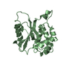
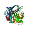
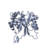
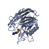
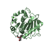
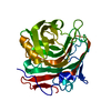
 Lactobacillus plantarum (bacteria)
 X-RAY DIFFRACTION /
  Authors
 Citation
 Structure visualization
 Downloads & links
 Other downloads
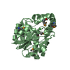


 PDBj
PDBj Assembly

 Mass: 22.990 Da / Num. of mol.: 2 / Source method: obtained synthetically / Formula: Na
 Mass: 18.015 Da / Num. of mol.: 154 / Source method: isolated from a natural source / Formula: H2O
 Sample preparation
 / Beamline: 19-ID / Wavelength: 0.9794 Å
 Processing